Beauty Through Medicine | Plastic Surgery & Cosmetology & Beauty Through Medicine & | Dr.Venkat Thota , Plastic Surgeon | Dr.Radha Thota, Medical Cosmetologist
When women consider breast reduction surgery, they often hesitate for two reasons. First, they worry about their weight (Body Mass Index or BMI). Second, they worry about their age.
A study from Baylor Scott & White Medical Center in Texas provides some clear answers.
The Study: 277 Women Analyzed
The researchers reviewed 277 breast reduction surgeries performed over a four-year period. They specifically looked at how age, weight, and the amount of tissue removed impacted the recovery process.
Here is what they found.
The Weight Factor: Slow Healing, Not Disaster
Patients often fear that a high BMI guarantees a surgical disaster. The data suggests otherwise.
The Good News: BMI was not associated with higher rates of major complications (like dangerous infections or blood clots).
The Reality: However, weight does matter for speed. The study found that women with a higher BMI were significantly more likely to require more than 2 months to heal.
Basically, heavier patients are safe, but they need more patience. The wounds may take longer to close completely.
The Age Factor: Minor Annoyances
Does age make surgery risky? Not exactly, but it does change the skin’s ability to bounce back.
The study found that greater age was linked to a higher rate of minor complications. These are usually superficial wound healing issues, like small scabs or separations along the incision line. They are annoying, but rarely dangerous.
Minor vs. Major Complications
It is important to understand what “complication” means in this context.
Minor Complications: These were common (49.1% of patients) and mostly involved superficial wounds. These heal with dressing changes and time.
Major Complications: These were rare (only 4.31%). No specific factor (age or weight) seemed to increase this risk.
The Bottom Line
This study offers reassurance. While having a higher BMI means you might need longer to heal, it does not necessarily rule you out for surgery.
As the authors conclude, the benefits of breast reduction—relief from back pain and improved quality of life—often outweigh the risks, even for selected patients with higher BMI.
If you smoke or have a higher Body Mass Index (BMI), finding a surgeon for breast reduction can be difficult. Many surgeons hesitate to operate on these “high-risk” patients.
The reason is simple: Blood Supply.
Nicotine shrinks blood vessels. This limits the oxygen reaching the healing tissue. In breast reduction surgery, this increases the risk of serious complications, specifically nipple necrosis (where the nipple tissue dies) or wound breakdown.
However, a study published in Plastic and Reconstructive Surgery presents a specialized technique designed to overcome these odds.
The Solution: The Three Dermoglandular Flap Technique
Surgeons from the University of Bari, Italy, developed a method specifically for challenging cases. They call it the Three Dermoglandular Flap technique.
How It Works
Standard reductions often rely on skin to hold the breast shape. Over time, skin stretches, and the breast droops again (recurrence).
This Italian technique uses a different approach:
Inferior-Central Pedicle: The surgeon keeps the nipple attached to a robust central bridge of tissue to ensure maximum blood flow.
Internal Support: They create three separate flaps of dermis (deep skin) and gland tissue.
The “Internal Bra”: These flaps are stitched together inside the breast. This creates a strong internal structure that supports the weight of the breast, independent of the skin.
The Test: Operating on “High-Risk” Patients
The researchers tested this method on the hardest-to-treat group. They selected 47 women who met strict criteria:
They were all smokers.
They had massive breasts (volume >1000 cc).
They had severe sagging (Grade 3 Ptosis).
They were overweight (Average BMI of 31.2).
The Results: Zero Nipple Loss
Given the high risks, the results were remarkable.
Safety: There were zero cases of partial or complete nipple necrosis.
Healing: There were zero cases of major wound breakdown.
Aesthetics: The technique produced a good cone shape with fullness in the upper breast.
Longevity: The results remained stable over time (up to 4 years of follow-up), proving the “internal support” worked.
What This Means for You
If you have been told you are “too high risk” for surgery due to smoking or weight, do not lose hope. While quitting smoking is always the best option for your health, surgical techniques exist that can handle challenging anatomy safely.
The Three Dermoglandular Flap technique offers a “safe and practical approach” for heavy, pendulous breasts. It prioritizes blood supply and structural support, ensuring you get the relief you need without the complications you fear.
When you consider breast reduction surgery, scarring is often a top concern. Traditionally, surgeons used the Wise-Pattern (or “Anchor”) technique. This leaves a scar around the areola, down the middle, and along the crease underneath the breast.
However, a newer technique called the Vertical Scar (or “Lollipop”) reduction has gained popularity. It eliminates the horizontal scar underneath the breast, leaving only a vertical line.
Patients often ask: Is the “Lollipop” technique just as safe? Can it handle large reductions? A study from the University of Illinois answers these questions.
The Study: Comparing 200 Breasts
Researchers wanted to compare these two popular methods directly. They conducted a “matched cohort study,” meaning they paired patients with similar ages and breast sizes to get a fair comparison.
Group A: 100 breasts operated on using the Superomedial Pedicle Vertical technique (“Lollipop”).
Group B: 100 breasts operated on using the Traditional Inferior Pedicle Wise-Pattern technique (“Anchor”).
This was a significant study because it looked at outcomes over a 3-year period.
The Results: Equal Safety, Less Scarring
The findings were reassuring for anyone hoping for fewer scars.
Large Reductions: The Vertical technique successfully handled large reductions. The average tissue removed was 815g (Vertical) versus 840g (Anchor) per breast.
Complications: There was no statistical difference in complications between the two groups. Healing issues and safety profiles were essentially the same.
Symptom Relief:100% of patients in both groups achieved relief from their symptoms (like back and neck pain).
The Verdict: The Vertical “Lollipop” reduction is a safe and effective alternative to the traditional “Anchor” method, even for larger breasts.
A Deeper Dive: It’s All About Blood Supply
In the accompanying discussion, expert surgeon Dr. Elizabeth Hall-Findlay highlights why the “Vertical” technique works so well. It often comes down to the “pedicle”—the bridge of tissue that carries blood to the nipple.
Dr. Hall-Findlay explains that the breast has four major arteries supplying it.
Inferior Pedicle (Traditional): Relies on the deep artery from the 4th interspace.
Medial/Superomedial Pedicle (Vertical): Relies on strong arteries from the 2nd or 3rd interspace.
She notes that a true Superomedial Pedicle is particularly robust. It incorporates the “very strong descending artery” from the second rib space. This provides a powerful “dual” blood supply, making it a reliable choice for keeping the nipple healthy during surgery.
Which Is Right for You?
This study proves that you do not necessarily need an “Anchor” scar just because you require a large reduction. The “Lollipop” technique offers excellent functional and aesthetic outcomes with a shorter scar.
However, anatomy varies. Your surgeon will choose the “pedicle” and scar pattern that best fits your specific blood supply and body shape.
We all love the latest technology. Whether it is a new iPhone or a high-tech kitchen appliance, we often assume “newer” means “better.” In plastic surgery, medical companies frequently market expensive new devices that promise faster recovery and less pain.
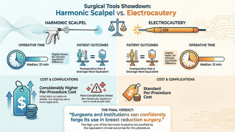
One such device is the Harmonic Scalpel. It claims to cut tissue and stop bleeding using ultrasonic vibrations rather than heat. But is it actually better than the standard tool surgeons have used for decades? A study from Dartmouth-Hitchcock Medical Center put this technology to the test.
The Tools: Electric vs. Ultrasonic
To understand the study, you must understand the tools:
Electrocautery (The Standard): This tool uses electricity to heat tissue. It cuts and seals blood vessels simultaneously. It is the gold standard for breast reduction.
Harmonic Scalpel (The Challenger): This device uses ultrasonic energy. It vibrates at high speeds to cut and coagulate tissue. It is often used in general surgery for procedures like thyroid removal.
The Experiment: A Side-by-Side Comparison
The researchers designed a clever study to remove outside factors like individual healing rates. They recruited 31 patients for bilateral breast reduction.
Here is the twist: They used both tools on the same patient.
One breast was operated on using the standard Electrocautery. The other breast was operated on using the Harmonic Scalpel. The assignment was random and blinded, meaning neither the patient nor the initial plan dictated which side got which tool.
The Results: Does Money Buy Better Results?
The study looked at three main things: speed, drainage (fluid buildup), and pain. The results might surprise you.
1. Speed (Operative Time)
The manufacturer claims the Harmonic Scalpel is more efficient. However, the study found the opposite. The median time for the Harmonic Scalpel was 33 minutes, compared to 31 minutes for standard electrocautery. While this difference was statistically significant, it is practically negligible. Basically, the fancy tool did not save time.
2. Pain and Drainage
Did the ultrasonic technology reduce pain or fluid buildup? No.
The researchers found no statistical difference in drainage volume or postoperative pain scores between the two sides. Patients felt the same, regardless of the tool used.
3. The Cost
This is the biggest difference. While the start-up costs for the machines were comparable, the per-procedure cost for the Harmonic Scalpel was considerably higher.
Expert Opinion: Why It Didn’t Work
In the accompanying discussion, Dr. Melissa Crosby from M.D. Anderson Cancer Center explains why this high-tech tool fell short.
The Harmonic Scalpel is excellent for surgeries like thyroidectomies because it replaces slow manual techniques like tying knots or using clips. However, in breast reduction, surgeons already use electrocautery for speed and sealing. Therefore, swapping it for the Harmonic device does not add efficiency; it just adds cost.
Dr. Crosby also noted that in an era of cost-effective medicine, we must critically appraise expensive gadgets to ensure they actually benefit the patient.
The Verdict
The study concludes that the Harmonic Scalpel is roughly equivalent to standard electrocautery—but it costs much more.
For patients, this is good news. You do not need to seek out a surgeon who uses this specific “high-tech” device to get a great result. The standard method is efficient, safe, and just as comfortable.
Frequently Asked Questions (FAQ)
Q: Does the Harmonic Scalpel reduce scarring?
A: This study did not find any advantage in terms of healing complications or drainage that would suggest better scarring. In fact, there were slightly more complications on the Harmonic side, though the number was too small to be certain.
Q: Why do some surgeons use it?
A: Some surgeons may prefer it for other types of surgery (like general surgery) and carry that preference over. However, evidence shows no specific benefit for breast reduction.
Q: Is Electrocautery safe?
A: Yes. It has been the standard in surgery for many years. It is effective at stopping bleeding (hemostasis) while cutting, which keeps the surgery safe and quick.
Women with extremely large breasts (Severe Gigantomastia) often face a difficult choice. They want relief from the heavy weight. However, surgeons often tell them they need a “Free Nipple Graft.”
This technique involves completely removing the nipple and sewing it back on as a skin graft. It is safe, but the trade-off is steep. Patients often lose nipple sensation, nipple projection, and the ability to breastfeed.
Why Is This Usually Necessary?
In standard breast reductions, surgeons rely on general anatomical rules to keep the nipple alive. We assume we know where the blood vessels are.
But in severe gigantomastia, the anatomy is stretched. The nipple sits very far away from the chest wall. Guessing the location of the blood vessels becomes risky. If the surgeon guesses wrong, the nipple could die. Therefore, many choose the Free Nipple Graft to be safe.
The Solution: Seeing Inside with Ultrasound
A study published in Plastic and Reconstructive Surgery offers a smarter, technology-driven alternative.
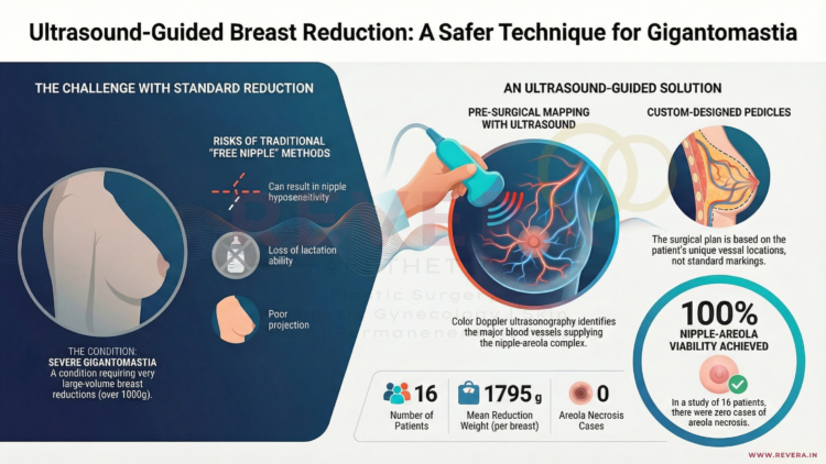
Researchers from Istanbul Medical Faculty decided not to guess. Instead, they used Color Doppler Ultrasonography before surgery to map out the breast.
How It Works
The Scan: Before the operation, the surgeon uses an ultrasound probe to scan the breast.
The Map: They identify exactly which blood vessels (perforators) are feeding the nipple-areola complex.
The Design: The surgeon designs a custom tissue bridge (“pedicle”) specifically for that patient. It protects those specific vessels.
Customizing the Surgery
The study showed that every woman is different.
Some patients had strong blood supply from the inner breast (Internal Mammary artery).
Others relied on the outer breast (Lateral Thoracic artery).
By using ultrasound, the surgeons could tailor the cut to the patient. They used superomedial, superolateral, or mediolateral designs depending on what the scan showed.
The Results: Safety Without Sacrifice
The study followed 16 patients with severe gigantomastia (average age 41).
Massive Reduction: The average weight removed was nearly 1.8 kg (about 4 lbs) per breast.
No Free Graft: None of the patients required a Free Nipple Graft.
What This Means for You
Having massive breasts does not mean you must sacrifice nipple sensation or shape. Technology can act as a “guide” for your surgeon.
If you are considering a significant reduction, ask your surgeon about preservation techniques. A personalized approach, guided by ultrasound, can offer the safety of a Free Nipple Graft without the downsides.
Breast reduction surgery is not just about making the breast smaller; it is about reshaping it to look proportional and centering the nipple on the new mound.
Most standard breast reduction techniques (like the popular Superomedial Pedicle) assume the nipple is starting from a standard sagging position. But what happens when a patient’s nipples are naturally positioned medially—meaning they sit closer to the cleavage or “point inward”?
In these cases, using standard techniques can be mechanically difficult. Trying to move an inward-facing nipple into a central position can restrict the blood supply or create tension, limiting how perfect the final result can be.
The Solution: The Superolateral Pedicle (SLP)
A new study published in Plastic and Reconstructive Surgery (August 2025) highlights a specific surgical approach designed exactly for this anatomy: the Superolateral Pedicle (SLP).
How It Works
In breast reduction, the nipple is kept alive on a “pedicle”—a bridge of tissue that preserves blood vessels and nerves.
Standard Way (Superomedial): The tissue bridge is usually kept on the inner/top side.
The SLP Way: For patients with medial nipples, the surgeon keeps the tissue bridge on the outer/top side (Superolateral).
By anchoring the nipple from the outside, the surgeon can more easily swing and rotate the nipple into the perfect central position without fighting the breast’s natural tissue resistance.
Is It Safe? The Research Say Yes
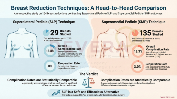
Researchers from MedStar Georgetown University Hospital in Washington, DC, reviewed 164 breast reductions to compare the safety of this specialized SLP technique against the standard method.
The Findings:
Comparable Safety: The complication rates were nearly identical between the two groups (13.8% for SLP vs. 13.3% for standard), proving that this technique is just as safe as the traditional method.
Effective Reduction: The technique worked well for significant reductions, with an average tissue removal of over 700g.
No Re-operations: In this specific study group, zero patients in the SLP group required a return to the operating room for complications, compared to 5 cases in the standard group.
Why This Matters For You
Anatomy is unique. If you have noticed that your nipples sit closer to your breastbone or point inward, standard techniques might not offer you the best aesthetic result. This research confirms that your surgeon has a validated, safe “tool in the toolkit” to correct medially positioned nipples and achieve a beautiful, centered look.
Frequently Asked Questions (FAQ)
Q: How do I know if I have “medially positioned” nipples?
A: If your nipples seem to sit closer to your cleavage rather than the center of your breast mound, or if they point inward towards each other, you likely have medial positioning. Your surgeon will assess this during your consultation.
Q: Does this technique leave different scars?
A: generally, no. The Superolateral Pedicle refers to the internal tissue handling. The external scars usually follow the standard “Wise Pattern” (Anchor) or Vertical (Lollipop) shape, just like a regular breast reduction.
Q: Is the recovery harder with this technique?
A: According to the study, complications such as wound healing issues (dehiscence) or fluid collection (seroma) were comparable to the standard technique, suggesting the recovery process is very similar.
Q: Can I still breastfeed with this technique?
A: Like the standard Superomedial technique, the SLP preserves a bridge of tissue carrying blood and nerve supply to the nipple. While breastfeeding can never be guaranteed after reduction surgery, techniques that preserve the pedicle generally offer a better chance than those that do not.
Breast reduction surgery (reduction mammaplasty) is one of the most life-changing procedures in plastic surgery, performed on over 100,000 patients annually. To ensure patients receive the safest and most effective care, the American Society of Plastic Surgeons (ASPS) convened a multidisciplinary work group to update their clinical practice guidelines.
Published in 2022, these guidelines reviewed thousands of studies to determine what truly works. Here is what the new evidence means for you as a patient.
1. It Is About Your Symptoms, Not the Scale
For years, insurance companies often demanded a specific weight of tissue be removed (e.g., 500g or 1000g) to qualify for coverage. The new guidelines challenge this outdated metric.
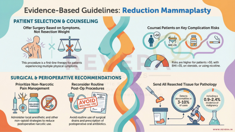
The ASPS now strongly recommends that surgery be offered as first-line therapy based on symptoms, not resection weight.
The Evidence: Studies show that relief from back pain, neck pain, and bra strap grooving is not correlated with the amount of tissue removed.
The Takeaway: If you have multiple physical symptoms (pain, rashes, grooving) that aren’t fixed by non-surgical methods, you are a candidate for surgery, regardless of whether you need a “small” or “large” reduction6666.
2. Drains Are No Longer Routine
One of the most dreaded parts of recovery for many patients is the use of surgical drains (tubes sticking out of the incision to collect fluid).
The guidelines bring good news: Plastic surgeons should not routinely use intraoperative drains for breast reduction patients.
Why? High-quality evidence shows no significant difference in complication rates (like hematomas) between patients with drains and those without.
The Benefit: avoiding drains means less discomfort during removal, lower costs, and less scarring.
3. Pain Management Has Evolved (Less Narcotics)
The modern approach to breast reduction focuses on multimodal pain managementto reduce the need for strong opioids (narcotics).
Local Anesthesia: The guidelines strongly recommend administering local anesthetic (numbing medication like lidocaine or bupivacaine) at the surgical site. This significantly improves pain scores immediately after surgery and reduces the time spent in the recovery room.
Non-Narcotic Strategies: Surgeons are encouraged to use non-opioid medications (such as Acetaminophen or NSAIDs) to manage pain safely.
4. Technique: The Pedicle Choice
The “pedicle” is the bridge of tissue that keeps your nipple alive and sensitive during the lift and reduction. The guidelines reviewed the two most common techniques:
Inferior Pedicle: The most commonly used, reliable for preserving blood supply.
Superomedial Pedicle: Preserves upper-pole fullness and avoids a long transverse scar.
The verdict? Both techniques are acceptable and effective. There is no significant difference in major complications between them, so your surgeon can choose the method best suited to your specific anatomy.
5. Important Risk Factors
To ensure safety, the guidelines identified specific factors that may increase the risk of complications. Patients should be counseled if they:
Are older than 50 years.
Have a Body Mass Index (BMI) greater than 35.
Use chronic corticosteroids.
Additionally, there is a strong recommendation regarding Nicotine: Patients identified as nicotine users should be referred to cessation programs and encouraged to stop smoking before surgery. Smoking significantly increases the risk of wound healing problems and infection.
6. Antibiotics and Pathology
Antibiotics: Extended courses of antibiotics after you go home are generally not recommended. A single dose given before surgery (within 1 hour of incision) is sufficient to prevent infection without causing antibiotic resistance.
Pathology: It is recommended that all breast tissue removed during the surgery be sent to the lab for evaluation to check for any hidden abnormal cells or high-risk lesions.
Frequently Asked Questions (FAQ)
Q: Do I have to try physical therapy before surgery?
A: The guidelines state that reduction mammaplasty should be offered as first-line therapy over non-operative treatments. There is no evidence that non-operative management (like special bras or therapy) provides effective long-term relief for breast hypertrophy.
Q: Will I have drains?
A: According to the 2022 guidelines, routine use of drains is not supported by evidence. However, exceptions may be made for specific high-risk cases or if liposuction is also performed.
Q: Does the “Pedicle” technique affect breastfeeding?
A: The guidelines noted that techniques preserving the subareolar parenchyma (the tissue under the nipple) increase the likelihood of breastfeeding success, but more research is needed to compare specific techniques directly.
For many women, breast reduction is about comfort and confidence. But for women with Gigantomastia (massive breasts), the condition is a severe medical burden.
Patients with massive breasts often suffer from debilitating back pain, skin infections, and significant mobility issues. Even more concerning, the sheer weight of the tissue can exacerbate pre-existing cardiac (heart) and respiratory (lung) problems.
For these patients, surgery is not a luxury—it is a necessity. However, because these patients often battle obesity or heart conditions, undergoing a long surgical procedure can be risky.
The Challenge: The 4-Hour Marathon
The most popular traditional methods for breast reduction (such as the McKissock or Robbins techniques) rely on complex internal sculpting. While effective, these surgeries typically require 3 to 4 hours of operative time.
For a patient with heart or lung issues, being under general anesthesia for four hours can be dangerous. Consequently, some women feel forced to consider a Total Mastectomy (complete removal of the breast) just to get relief, or they avoid surgery altogether.
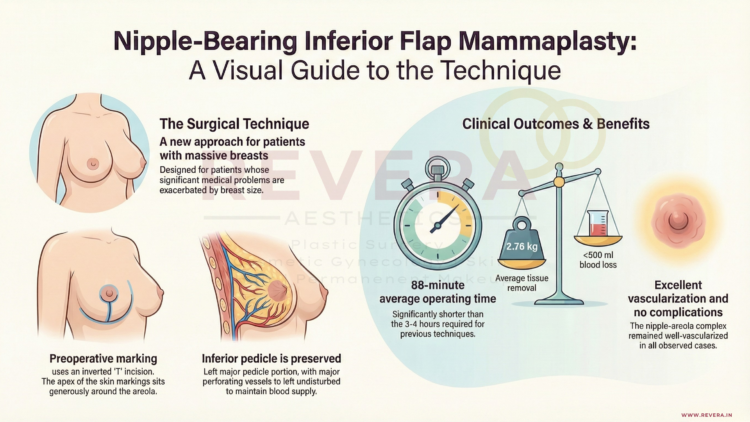
The Solution: Nipple-Bearing Inferior Flap Mammaplasty
A technique published in Plastic and Reconstructive Surgery offers a powerful solution specifically designed for massive weight reduction with maximum safety.
Developed by a surgical team in Preston, England, this technique focuses on speed and blood supply safety.
How It Works
Unlike complex sculpting methods that require separating the breast tissue from the chest wall (“undermining”), this technique leaves the base of the breast undisturbed.
Wide Base: The surgeon creates a wide, supportive base of tissue (the “inferior flap”) that keeps the nipple attached to its natural blood supply.
No Grafting Needed: Unlike some rapid reduction techniques that cut the nipple off and sew it back on as a skin graft, this method keeps the nipple connected, preserving its vitality.
Efficient Removal: A wedge of tissue is removed down to the pectoral fascia, and the remaining flaps are brought together securely.
The Results: Faster and Safer
The study followed patients with massive breasts and significant medical problems who underwent this specific procedure. The results were transformative:
Drastically Reduced Surgery Time: The average operation took only 88 minutes (compared to the standard 3–4 hours).
Massive Weight Loss: The average tissue removal was 2.76 kg (over 6 lbs) per patient.
Safety: There was minimal blood loss (less than 500 mL on average).
Nipple Survival: Because the nipple was carried on a wide, robust flap, it remained well-vascularized with no complications in the study group.
Is This Right For You?
This technique is ideal for women who:
Have extremely large breasts causing medical distress.
Have been told they are “high risk” for long surgeries due to obesity or heart/lung conditions.
Want significant size reduction without resorting to a total mastectomy.
We believe that health and mobility should never be out of reach. If you are suffering from the weight of massive breasts, contact us to discuss if this rapid-reduction technique is an option for you.
Frequently Asked Questions (FAQ)
Q: Will I lose my nipple sensation?A: This technique is a “Nipple-Bearing” procedure. Unlike a “Free Nipple Graft” (where the nipple is completely detached), this method maintains a wide bridge of tissue connecting the nipple to the body’s blood and nerve supply, which helps preserve the nipple-areola complex.
Q: How are the scars placed?A: The incision design usually results in an inverted-U or inverted-L shape on the lower breast. While scarring is inevitable in reduction surgery, the primary goal of this specific technique is massive volume reduction to improve heart and lung health.
Q: Why is a shorter surgery better?A: For patients with pre-existing medical conditions (like high blood pressure, asthma, or obesity), prolonged anesthesia increases the risk of complications such as clots (DVT) or respiratory distress. Reducing the time from 4 hours to roughly 90 minutes significantly lowers this risk.
For millions of women undergoing mastectomy (breast removal) for cancer treatment or prevention, the primary focus is survival. The secondary focus is often reconstruction—restoring the physical appearance of the breast.
However, there is a “silent” side effect that is rarely discussed but deeply felt: Numbness.
During a mastectomy, the sensory nerves that provide feeling to the breast skin and nipple are often cut.
A Little Background on Anatomy
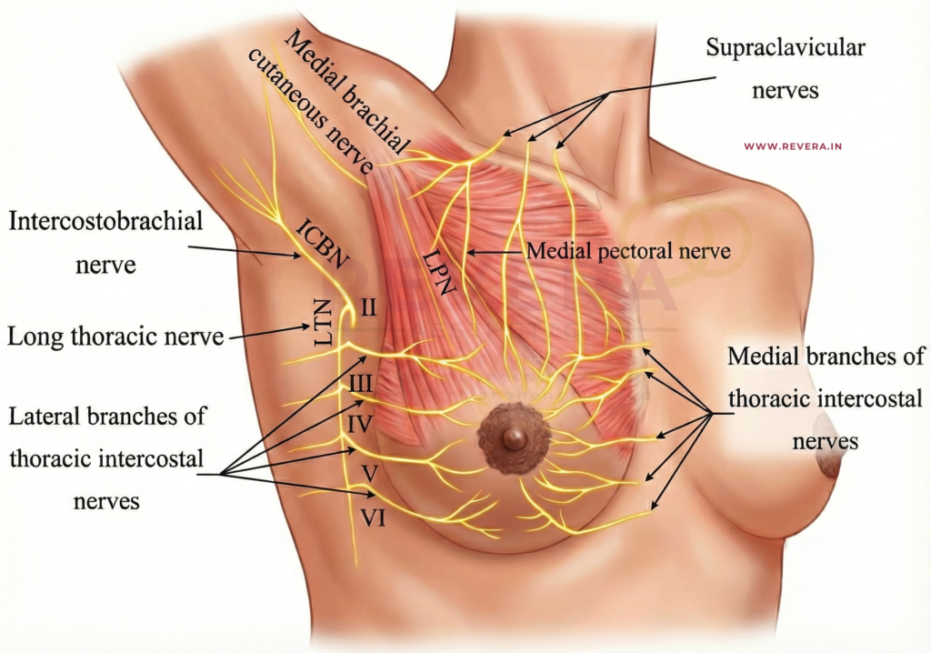
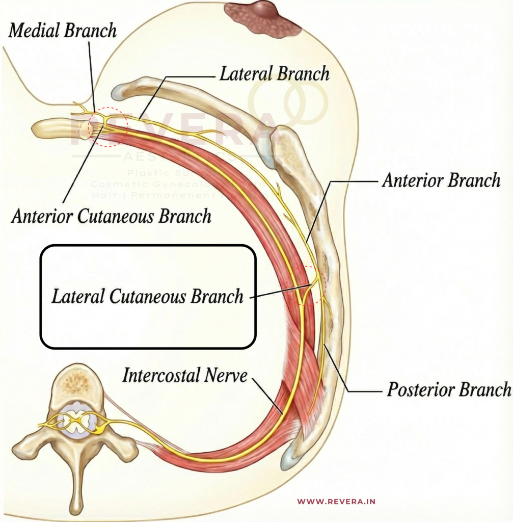
To understand why numbness occurs, we must look at how the breast receives feeling. The sensory innervation to the breast originates from the medial and lateral cutaneous branches of the third to fifth intercostal nerves.
Shutterstock
The Path of the Nerves: The third, fourth, and fifth intercostal nerves give off lateral cutaneous branches that pierce the chest wall (near the mid-axillary line, or the side of your rib cage). These branches divide into anterior and posterior parts, supplying the skin of the side and front of the chest.
The Anterior Branches: The anterior cutaneous branches (the terminal parts of these nerves) supply the skin on the chest wall toward the center (sternum).
The 3rd nerve covers the upper-mid chest.
The 4th nerve covers the central chest and the medial (inner) breast area – The Nipple Line.
The 5th nerve covers the lower-mid chest and the inframammary fold (where the breast meets the ribs).
These nerves provide sensory innervation (known as dermatomes) to specific chest and abdominal areas and are crucial for sensation and pain management (such as nerve blocks).
For years, women have accepted that their reconstructed breasts, while looking beautiful, would permanently feel numb—like “wearing a bra made of your own skin.”
The Missing Link: Finding the Nerve
Restoring sensation (Resensation) is possible. Surgeons can perform a “nerve transfer,” connecting a nerve from your chest wall to the nerves in the reconstructed breast.
So, why isn’t this done all the time?
One major hurdle has been the difficulty of finding the right donor nerve. The human body is complex, and searching for a tiny nerve (often only 2 millimeters wide) during a long surgery can be like finding a needle in a haystack.
The Breakthrough: A Roadmap for Sensation
A pivotal study published in Plastic and Reconstructive Surgery has provided surgeons with a reliable “treasure map” to find these elusive nerves.
Research conducted by Dr. Rebecca Knackstedt and Dr. Risal Djohan (along with their team in Cleveland and Toledo, Ohio), utilized precise anatomical studies to identify the exact hiding place of the nerve responsible for breast sensation.
What They Found
The researchers discovered that the Lateral Intercostal Branch (the nerve key to breast feeling) is located in a highly predictable spot:
It almost always exits from under the 4th Rib.
It sits consistently near the edge of the Pectoralis Minor muscle.
It travels safely underneath the thoracodorsal vessels (major blood vessels in the armpit area).
Why This Matters for Your Surgery
This study transforms a “search mission” into a precise procedure. Because surgeons now know exactly where to look, we can locate the nerve with much greater accuracy. The study identified the nerve’s location as:
10 to 15 cm from the sternum (breastbone).
8 to 16 cm from the mid-clavicular line.
Near the lateral border of the armpit muscle (Pectoralis minor) or within 2 cm from it.
By using these precise coordinates, surgeons can:
Locate the nerve quickly, reducing surgery time.
Preserve the nerve more effectively.
Perform Nerve Allografts: Connect this sensation-carrying nerve to your reconstructed breast tissue using a nerve graft.
Moving Beyond “Looking Normal”
We believe that feeling whole means more than just looking in the mirror; it’s about feeling a hug, noticing a change in temperature, and reclaiming your body’s sensation.
Thanks to anatomical breakthroughs like this study from Ohio, Breast Neurotization (nerve repair) is becoming a more standard and successful part of breast reconstruction.
Frequently Asked Questions (FAQ)
Q: If I have this procedure, will my sensation be 100% normal?
A: “Normal” is a strong word. Nerve regeneration is slow and complex. Most patients do not regain perfect, pre-surgery sensitivity. However, the goal is to transition from “numbness” to “protective sensation” (feeling touch and pressure) and, in many cases, erogenous sensation. It is a vast improvement over having no feeling at all.
Q: Does this add time to the surgery?
A: Yes, nerve repair does add some time to the reconstruction surgery. However, thanks to the “mapping” provided by this research, the time taken to find the nerve is significantly reduced, making the addition of nerve repair much more feasible.
Q: Can this be done if I had a mastectomy years ago?
A: Breast reinnervation is most successful when performed at the same time as the mastectomy (Immediate Reconstruction). Doing it years later is much more difficult because the nerve endings may have scar tissue or have become dormant. However, it is always worth discussing with your surgeon.
Q: Is this only for implant reconstruction or flap reconstruction?
A: Nerve grafts can be used in both. In DIEP Flap (using your own tissue), surgeons connect the chest nerve to the nerve in the tummy tissue. In Implant reconstruction, the nerve is connected to the remaining skin or nipple nerves.
Reference
Knackstedt, Rebecca M.D., Ph.D.; Gatherwright, James M.D.; Cakmakoglu, Cagri M.D.; Djohan, Michelle M.S.; Djohan, Risal M.D. “Predictable Location of Breast Sensory Nerves for Breast Reinnervation.” Plastic and Reconstructive Surgery. February 2019. Cleveland Clinic & University of Toledo, Ohio.
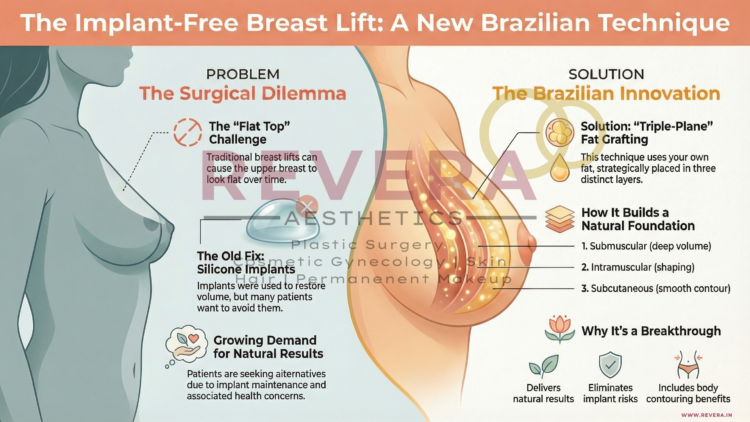
However, there has always been a surgical dilemma. Traditional lifts are excellent at removing excess skin and reshaping the breast, but they often struggle to maintain “upper pole projection”—that desirable fullness at the top of the breast (the décolletage). Over time, gravity can cause the breast tissue to settle, sometimes leaving the upper breast looking flat.
Historically, the solution to this was using silicone implants to fill that upper volume. But what if you don’t want implants?
The “Implant-Free” Movement
While implants remain a popular choice, many of our patients are seeking natural alternatives. Some wish to avoid the maintenance of implants, while others are concerned about rare but documented risks associated with silicone, such as Breast Implant-Associated Anaplastic Large-Cell Lymphoma (BIA-ALCL) or autoimmune symptoms.
If you desire fullness and lift but want to stay 100% natural, a groundbreaking new technique known as Triple-Plane Autologous Fat Grafting might be the answer.
Innovative Research from Brazil
We pride ourselves on staying up-to-date with global advancements in aesthetic medicine. This specific technique comes from a cutting-edge study titled “Breast codes: triple plane autologous fat grafting,” published in August 2025 in the prestigious Plastic and Reconstructive Surgery journal.
The study was conducted by Dr. Ricardo T. Nóra, a plastic surgeon based in Sinop, Mato Grosso, Brazil, in collaboration with Dr. Lydia M. Ferreira from the Division of Plastic Surgery at the Federal University of São Paulo, Brazil.
Brazil has long been recognized as a world leader in cosmetic surgery innovation. In this study, Dr. Nóra and Dr. Ferreira standardized a method to enhance breast shape without foreign bodies, addressing the limitations of previous techniques.
What is the “Triple-Plane” Technique?
Autologous Fat Graftinginvolves taking fat from an area where you have a little extra (like the abdomen or thighs) and transferring it to the breasts.
The innovation introduced by Dr. Nóra and Dr. Ferreira is the “Triple-Plane” approach. Instead of randomly injecting fat, this technique systematically places fat in three specific layers (planes) to build a lasting structural foundation:
Submuscular: Deep placement under the muscle for foundational volume.
Intramuscular: Placement within the muscle for dynamic shaping.
Subcutaneous: Placement just under the skin for smooth contouring and softness.
Why This Matters for You
This innovative approach addresses the “flat top” issue without requiring a foreign object in your body. By strategically layering the fat according to these new “Breast Codes,” surgeons can now create that coveted upper-breast fullness during a lift or reduction.
Key Benefits:
Natural Results: Uses your own tissue, so the look and feel are entirely you.
Safety: Eliminates the risks associated with long-term silicone implants.
Body Contouring: You get the added benefit of liposuction in the donor area.
Scientifically Backed: Based on rigorous surgical protocols developed by leading experts in Brazil.
Experience the Future of Breast Surgery
If you are looking for a natural, long-lasting enhancement, let’s discuss if this technique is right for your body goals. Contact our clinic today at +919866224871 to schedule a consultation. Chat with us on WhatsApp.