Young woman and surgeon giving thumbs up during a breast reduction consultation for the omega resection technique. Best Plastic Surgeon in Hyderabad
Written by revera-admin

The Omega Resection: A Faster, Safer Alternative for Breast Reduction

The Search for a Simpler Technique

Symptomatic breast hypertrophy causes substantial physical distress, forcing millions of women to live with chronic back, neck, and shoulder pain. While conservative measures like physical therapy or weight loss rarely provide lasting relief, reduction mammoplasty consistently delivers high patient satisfaction. Consequently, it remains the gold standard treatment for macromastia. The Omega Resection around the NAC is A Faster, Safer Alternative for Breast Reduction

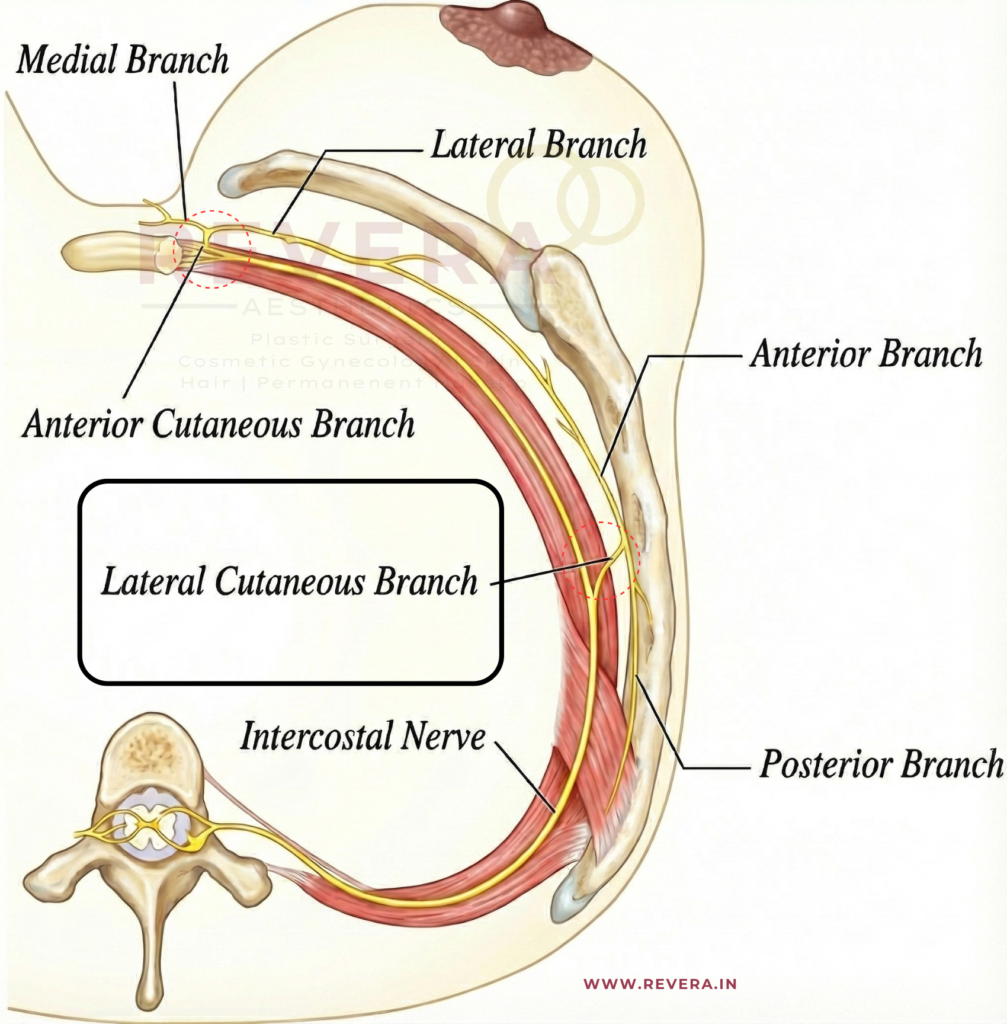
Although the traditional inferior pedicle approach is highly popular among plastic surgeons, it can be technically complex and time-consuming. To simplify this process, a team of Swiss plastic surgeons evaluated a modified approach: The Omega Resection Pattern Technique.

The Anatomy of the Omega Cut

The omega technique relies entirely on the stable blood supply of a standard inferior pedicle. However, it radically alters how the surgeon resects the tissue.

How the Surgical Technique Works:

  1. The Markings: First, the surgeon draws an omega-shaped (Ω) outline around the nipple-areola complex (NAC) while the patient stands upright.
  2. The Incision: Subsequently, the surgeon makes a precise incision that extends straight down to the pectoralis fascia.
  3. The Resection: Most importantly, instead of shaving away tissue piecemeal, the surgeon removes the deep breast parenchyma en bloc from both sides of the pedicle simultaneously.
  4. The Closure: Finally, the surgeon shapes the remaining tissue into a tight, natural contour and closes the skin using a tension-free, traditional inverted-T pattern.

Key Findings: Shorter OR Times and Lower Complications

The retrospective review analyzed 67 reduction mammoplasties performed over a 10-year period by a single senior plastic surgeon. The average amount of tissue resected was 826 grams per breast, with cases reaching up to 2,307 grams.

The Omega Resection around the NAC is A Faster, Safer Alternative for Breast Reduction while still using the standard inferior pedicle.

When the researchers compared their results to classic literature, the data revealed two distinct advantages:

1. Significantly Faster Operation Times

The mean operation time for the omega technique alone was 149 minutes. Because the en bloc resection eliminates the need for tedious, incremental tissue trimming and extensive dermal de-epithelialization, this technique is statistically faster than standard alternatives. Specifically, it outperforms the traditional inferior pedicle (177 minutes) and the superior pedicle approach (166 minutes).

2. A Minimal Complication Rate

The overall complication rate was 15%, and notably, zero major complications occurred during the 12-month follow-up window.

   [Surgical Technique] ───► Total Complication Rates
              │
              ├──► Traditional Inferior Pedicle: 29.7%
              ├──► Traditional Superior Pedicle: 19.6%
              ├──► Superomedial Pedicle: 16.9%
              └─► OMEGA RESECTION PATTERN: 15.0% (All Minor)

Minor complications included limited wound dehiscence (9%), minor surgical site infections (4.5%), and a single postoperative hematoma (1.5%). Furthermore, despite performing massive resections on patients with severe ptosis, no cases of full or partial nipple necrosis occurred.

Predicting Surgical Success: The Protective Factors

Through univariate logistic regression analysis, the authors identified several patient characteristics and clinical decisions that significantly diminished the risk of postoperative complications.

  • Patient Biology: Maintaining a normal BMI and a non-smoker status served as powerful protective factors against delayed wound healing. Indeed, out of the seven active smokers in the study, four developed wound complications.
  • Breast Anatomy: Resection weights between 500 and 1,500 grams and a sternal notch-to-nipple (NTN) distance under 30 cm were statistically safer.
  • Clinical Care: Keeping a patient overnight for inpatient hospitalization, avoiding multiple simultaneous surgeries (like combining the reduction with an abdominoplasty), and removing surgical drains at least one day after surgery all predicted a lower incidence of complications.

Conclusion

The 10-year review demonstrates that the omega resection pattern technique is an effective, safe, and exceptionally fast option for treating bilateral macromastia. Additionally, it serves as an excellent tool for unilateral contralateral breast symmetrisation following breast cancer surgery. By streamlining the resection into a single, predictable block, it offers a highly dependable alternative for modern aesthetic and reconstructive practices.

Frequently Asked Questions (FAQ)

Q: What exactly is an “en bloc” resection? A: En bloc means removing tissue as a single, whole piece rather than cutting it away in small fragments. In this technique, the entire excess outer section of the breast is removed in one unified block, which saves significant operative time.

Q: Can this technique be used on massive breasts or gigantomastia? A: Yes. The study included patients who required resections of over 2,000 grams per breast, proving the technique is safe even for massive tissue removal.

Q: Why does standard inferior pedicle surgery have a higher complication rate? A: Traditional approaches often involve extensive undermining and skin reshaping, which puts stress on the incision lines. Because the omega pattern cuts cleanly to the chest wall, it reduces tissue trauma and handles tension efficiently.

Reference

Written by revera-admin

Your Post-Breast Reduction Surgery Mammogram: What Has Changed?

The New “Baseline”

If you have a breast reduction, your breasts undergo significant internal changes. It is not just the outside that is reshaped; the internal glandular tissue is moved, stitched, and sometimes combined with liposuction.

Because of this, your mammograms will look different for the rest of your life. Dr. Joan Robertson conducted a study on 50 patients to document these “constant and significant” changes.

Why Does the Image Change?

The surgery involves moving the “nipple-areola complex” and the underlying tissue. This shifting creates specific features that a radiologist (a doctor who reads X-rays) must recognize so they don’t mistake them for something more serious.

1. Internal Scarring (Fibrosis)

As the breast heals, internal “scar tissue” forms where the incisions were made. On a mammogram, this can appear as thickened areas or shadows.

2. Oil Cysts and Calcifications

Sometimes, small areas of fat tissue lose their blood supply during surgery. This is called fat necrosis. While harmless, it can turn into “oil cysts” or tiny calcium deposits (calcifications) that show up clearly on an X-ray.

3. The “Mediolateral” Shift

Interestingly, Dr. Robertson found that these changes are often most visible on the mediolateral view (the side-to-side view) rather than the cranio-caudad (top-down) view.

Safety First: The Hidden Findings

As we discussed in the Emory University study, surgeons always send the removed tissue to a lab to check for hidden abnormalities.

  • The Benefit: This routine check catches hidden findings in 1.8% of general patients.
  • The Protocol: Having a “normal” mammogram before surgery is standard, but the lab test after surgery is an extra layer of safety.

Tips for Your Future Mammograms

  1. Wait for the Settling: Most surgeons recommend waiting 3 to 6 months after surgery before getting a new “baseline” mammogram.
  2. Inform the Tech: Always tell the mammogram technician that you have had a breast reduction. They will place specialized “scar markers” (tiny stickers) on your skin so the radiologist knows where the surgical lines are.
  3. Provide Old Films: If possible, give your radiologist your mammograms from before the surgery. Comparing the “old” breast to the “new” breast helps them identify which changes are purely surgical.

Conclusion

A breast reduction does not make it harder to detect cancer, but it does change the “landscape” of your breast tissue. By understanding these changes and communicating with your medical team, you can continue your routine screenings with confidence and peace of mind.


Ask yourself “Who is the Best Plastic Surgeon Near Me?”.

Revera Clinic caters with the Best Plastic Surgeon in Hyderabad!

Breast Reduction Surgery Cost varies between individuals!

Contact us to know if you are a suitable candidate for Breast Reduction Surgery!


Frequently Asked Questions (FAQ)

Q: Does breast reduction increase the risk of breast cancer?

A: No. In fact, some studies suggest that because you have less breast tissue after surgery, the overall risk may slightly decrease.

Q: What if my mammogram shows “calcifications”?

A: Post-surgical calcifications are very common. Radiologists can usually tell the difference between “benign” (harmless) surgical calcifications and those that require further testing.

Q: Should I get a mammogram right before my surgery?

A: Yes. The ASPS Guidelines recommend a preoperative mammogram for most women over the age of 35 or those with a family history of breast cancer.


References

Written by revera-admin

Breast Reduction: Is It Safe if You are Morbidly Obese?

The Weight Barrier

Many plastic surgeons hesitate to perform breast reductions on patients with a high Body Mass Index (BMI). This is especially true for “morbidly obese” patients (BMI of 40 or higher).

Surgeons often worry about a higher risk of infections and poor wound healing. They also fear the challenges of Gigantomastia. This is when a surgeon must remove more than 2,000 grams of tissue from each breast.

But does the data actually support turning these patients away? A study published in Plastic and Reconstructive Surgery investigated this exact question.

The Study: Analyzing 179 Patients

Researchers in Galveston, Texas, performed a retrospective review of 179 patients. They wanted to see which factors truly caused complications. They looked at:

  • Body Mass Index (BMI).
  • The weight of the tissue removed.
  • The patient’s age.
  • Smoking status.
  • Other health conditions (comorbidities).

The Findings: Safety Across the Scale

The researchers found an overall complication rate of 50%. While this number seems high, it mostly consisted of minor healing issues common in large-volume surgeries.

Crucially, the study found no statistical difference in complications based on:

  1. BMI: Patients with a BMI over 40 were just as safe as those with lower BMIs.
  2. Reduction Size: Removing massive amounts of tissue (>2000g) did not increase the danger.
  3. Age: Older patients did not face more risks than younger ones in this group.

Furthermore, smoking status and other medical conditions did not significantly impact the complication rates in this study.

The Conclusion: A Green Light for Surgery

The study reached a bold conclusion. It is as safe to perform large-volume breast reductions in morbidly obese patients as it is in anyone else.

What This Means for You

If you have a high BMI and suffer from the weight of very large breasts, you may have been told to “lose weight first.” While losing weight is generally healthy, this research proves that you do not have to wait to find relief from physical pain.

Modern guidelines from the American Society of Plastic Surgeons (ASPS) agree. They recommend that surgery be offered based on your symptoms, not just your weight.

If you suffer from back pain, rashes, or shoulder grooving, you deserve a consultation. A skilled surgeon can perform your reduction safely, regardless of your starting weight.

—————

Ask yourself “Who is the Best Plastic Surgeon Near Me?”.
Contact us to know if you are a suitable candidate for Breast Reduction.

Revera Clinic caters with the Best Plastic Surgeon in Hyderabad!


Frequently Asked Questions (FAQ)

Q: Will a high BMI make my recovery longer?

A: Possibly. While major complications are not higher, some studies suggest that patients with a BMI over 35 may take longer than two months to heal completely.

Q: What is “Gigantomastia”?

A: This is a medical term for extremely large breasts. It usually applies when a surgeon needs to remove more than 2,000 grams (about 4.4 lbs) from each breast.

Q: Are certain surgical techniques safer for obese patients?

A: This study looked at various methods, including inferior pedicles and free nipple grafts. It found that the specific surgical method did not change the complication rate.


References

  • [1] Roehl, Kendall M.D.; et al. “Breast Reduction: Safe in the Morbidly Obese?” Plastic and Reconstructive Surgery 122(2):p 370-378, August 2008.
  • [2] Perdikis, Galen M.D.; et al. “Evidence-Based Clinical Practice Guideline: Revision: Reduction Mammaplasty.” Plastic and Reconstructive Surgery 149(3):p 392e-409e, March 2022.
Written by revera-admin

Hidden Findings: Why We Test Tissue After Breast Reduction

The Routine Lab Test

When you undergo a breast reduction, your surgeon removes excess fat, skin, and glandular tissue. But what happens to that tissue?

Most patients assume it is simply discarded. However, standard safety protocols require this tissue to be sent to a lab for evaluation. While the goal of surgery is relief from physical pain, this routine step can sometimes uncover “occult” (hidden) medical findings.

The Evidence: The ASPS Recommendation

The American Society of Plastic Surgeons (ASPS) provides clear “Gold Standard” guidelines for this procedure.

  • The Rule: Plastic surgeons should send breast tissue from all patients for pathologic evaluation.
  • The Benefit: This allows for the early detection of cancer or high-risk lesions.
  • The Rationale: Preoperative mammograms are helpful but not perfect. They do not always catch the tiny abnormalities found in surgical specimens.

What the Research Says: The Emory University Study

A major study from Emory University analyzed the records of 1,014 patients who had breast reductions over 20 years. The researchers wanted to know how often hidden (occult) cancer or high-risk cells were found.

They split the patients into two groups:

  • Group A: Women with no history of breast cancer.
  • Group B: Women who had a previous breast cancer diagnosis.

The Incidence of Hidden Findings

The study found that these hidden findings are “not uncommon”.

  1. For General Patients (Group A): High-risk or malignant cells were found in 1.8% of patients.
  2. For Cancer Survivors (Group B): The risk was much higher, 8% of these patients had hidden findings in their reduction specimens.

Who Is at Higher Risk?

The research identified two major “positive predictors” for finding hidden abnormalities during surgery:

  • Increasing Age: As patients get older, the likelihood of a positive finding increases.
  • Personal History: Having a previous breast cancer diagnosis is a significant risk factor.

Why This Matters for Your Safety

Finding these cells early is a major benefit. Identifying high-risk lesions allows your medical team to start early treatment or more frequent screening.

As the authors of the study concluded, it is crucial for surgeons to maintain open communication with the lab. This ensures that if something is found, your follow-up care is precise and effective.


Frequently Asked Questions (FAQ)

Q: If my mammogram was clear, do I still need a lab test?

A: Yes. Research shows that 81% of patients with abnormal findings in their surgery tissue had a “normal” preoperative mammogram. The lab test is a necessary safety net.

Q: Is it common to find cancer during a breast reduction?

A: No, it is rare. In general patients, the risk is about 1.8%. However, “high-risk” cells (which are not cancer but could lead to it) are found slightly more often.

Q: Does insurance cover the cost of the lab test?

A: Generally, because this is a standard-of-care recommendation from the ASPS, it is treated as a necessary part of the medical procedure.


References

  • [1] Razavi, Seyed Amirhossein M.D.; et al. “The Incidence of Occult Malignant and High-Risk Pathologic Findings in Breast Reduction Specimens.” Plastic and Reconstructive Surgery 148(4):p 534e-539e, October 2021.
  • [2] Perdikis, Galen M.D.; et al. “Evidence-Based Clinical Practice Guideline: Revision: Reduction Mammaplasty.” Plastic and Reconstructive Surgery 149(3):p 392e-409e, March 2022.
Written by revera-admin

Can a “Heavy Chest” Actually Hurt Your Lungs?

The Weight on Your Chest

Women with macromastia (excessively large breasts) often complain of a “heavy” feeling on their chest. They may feel short of breath during exercise or even while resting.

Is this just a sensation? Or does the weight of the breasts actually restrict the lungs from filling with air?

A Randomized Controlled Trial from the University of Hull in the United Kingdom sought to answer this medical question.

The Study: Testing Lung Capacity

The researchers wanted to see if removing the breast weight improved lung function physically. They conducted a high-quality study (Randomized Controlled Trial) with 73 women.

  • Group 1 (Surgery): These women had breast reduction surgery immediately (within 6 weeks).
  • Group 2 (Control): These women waited 6 months before having surgery.

The doctors used spirometry (lung function tests) to measure how much air the women could inhale and exhale. They compared the results between the two groups.

The Findings: Weight Matters

The results revealed an interesting connection between breast size and breathing.

When they looked at the groups as a whole, the difference wasn’t immediately obvious. However, when they looked closer at the surgery group, they found a clear pattern.

  • The Correlation: There was a positive correlation between the weight of the tissue removed and the improvement in lung function.
  • The Takeaway: The more weight the surgeon removed, the better the patient’s lung test scores (like Peak Expiratory Flow) became.
  • Significant Improvement: Specifically, the study showed a significant improvement in Forced Vital Capacity (FVC). This measures the total amount of air you can forcibly exhale from your lungs.

Why Does This Happen?

Large breasts can act like a physical weight on the chest wall. This external weight may prevent the ribcage from expanding fully when you take a deep breath.

By performing a reduction mammaplasty, the surgeon removes this restriction. This allows the chest wall to move more freely, helping the lungs expand to their full predicted capacity.

Conclusion

If you have massive breasts and struggle to catch your breath, it might not just be “in your head.” It could be a mechanical restriction.

This study confirms that for women with heavy breasts, reduction surgery does more than relieve back pain. It correlates with a measurable improvement in pulmonary (lung) function.


Reference

[1] Iwuagwu, Obi C. F.R.C.S.; et al. “Does Reduction Mammaplasty Improve Lung Function Test in Women with Macromastia? Results of a Randomized Controlled Trial.Plastic and Reconstructive Surgery 118(1):p 1-6, July 2006.

Written by revera-admin

A Safer Breast Reduction for Smokers? The “Three Flap” TechniqueThe Risk: Why Surgeons Turn Smokers Away

The Risk: Why Surgeons Turn Smokers Away

If you smoke or have a higher Body Mass Index (BMI), finding a surgeon for breast reduction can be difficult. Many surgeons hesitate to operate on these “high-risk” patients.

The reason is simple: Blood Supply.

Nicotine shrinks blood vessels. This limits the oxygen reaching the healing tissue. In breast reduction surgery, this increases the risk of serious complications, specifically nipple necrosis (where the nipple tissue dies) or wound breakdown.

However, a study published in Plastic and Reconstructive Surgery presents a specialized technique designed to overcome these odds.

The Solution: The Three Dermoglandular Flap Technique

Surgeons from the University of Bari, Italy, developed a method specifically for challenging cases. They call it the Three Dermoglandular Flap technique.

How It Works

Standard reductions often rely on skin to hold the breast shape. Over time, skin stretches, and the breast droops again (recurrence).

This Italian technique uses a different approach:

  1. Inferior-Central Pedicle: The surgeon keeps the nipple attached to a robust central bridge of tissue to ensure maximum blood flow.
  2. Internal Support: They create three separate flaps of dermis (deep skin) and gland tissue.
  3. The “Internal Bra”: These flaps are stitched together inside the breast. This creates a strong internal structure that supports the weight of the breast, independent of the skin.

The Test: Operating on “High-Risk” Patients

The researchers tested this method on the hardest-to-treat group. They selected 47 women who met strict criteria:

  • They were all smokers.
  • They had massive breasts (volume >1000 cc).
  • They had severe sagging (Grade 3 Ptosis).
  • They were overweight (Average BMI of 31.2).

The Results: Zero Nipple Loss

Given the high risks, the results were remarkable.

  • Safety: There were zero cases of partial or complete nipple necrosis.
  • Healing: There were zero cases of major wound breakdown.
  • Aesthetics: The technique produced a good cone shape with fullness in the upper breast.
  • Longevity: The results remained stable over time (up to 4 years of follow-up), proving the “internal support” worked.

What This Means for You

If you have been told you are “too high risk” for surgery due to smoking or weight, do not lose hope. While quitting smoking is always the best option for your health, surgical techniques exist that can handle challenging anatomy safely.

The Three Dermoglandular Flap technique offers a “safe and practical approach” for heavy, pendulous breasts. It prioritizes blood supply and structural support, ensuring you get the relief you need without the complications you fear.


Reference

[1] Pascone, Michele M.D.; Di Candia, Michele M.D.; Pascone, Christian M.D. “The Three Dermoglandular Flap Support in Reduction Mammaplasty.Plastic and Reconstructive Surgery 130(1):p 1e-10e, July 2012.


Social Media Hashtags

#BreastReduction #PlasticSurgerySafety #SmokersSurgery #HighBMISurgery #BreastLift #MedicalResearch #SurgicalInnovation #MassiveWeightLoss #ReconstructiveSurgery #PatientEducation

Infographic showing advantages for going for USG mapping of Blood vessels supplying the Breast
Written by revera-admin

Massive Breast Reduction: How Ultrasound Can Save Your Nipple Sensation

The Fear of the “Free Nipple Graft”

Women with extremely large breasts (Severe Gigantomastia) often face a difficult choice. They want relief from the heavy weight. However, surgeons often tell them they need a “Free Nipple Graft.”

This technique involves completely removing the nipple and sewing it back on as a skin graft. It is safe, but the trade-off is steep. Patients often lose nipple sensation, nipple projection, and the ability to breastfeed.

Why Is This Usually Necessary?

In standard breast reductions, surgeons rely on general anatomical rules to keep the nipple alive. We assume we know where the blood vessels are.

But in severe gigantomastia, the anatomy is stretched. The nipple sits very far away from the chest wall. Guessing the location of the blood vessels becomes risky. If the surgeon guesses wrong, the nipple could die. Therefore, many choose the Free Nipple Graft to be safe.

The Solution: Seeing Inside with Ultrasound

A study published in Plastic and Reconstructive Surgery offers a smarter, technology-driven alternative.

Researchers from Istanbul Medical Faculty decided not to guess. Instead, they used Color Doppler Ultrasonography before surgery to map out the breast.

How It Works

  1. The Scan: Before the operation, the surgeon uses an ultrasound probe to scan the breast.
  2. The Map: They identify exactly which blood vessels (perforators) are feeding the nipple-areola complex.
  3. The Design: The surgeon designs a custom tissue bridge (“pedicle”) specifically for that patient. It protects those specific vessels.

Customizing the Surgery

The study showed that every woman is different.

  • Some patients had strong blood supply from the inner breast (Internal Mammary artery).
  • Others relied on the outer breast (Lateral Thoracic artery).

By using ultrasound, the surgeons could tailor the cut to the patient. They used superomedial, superolateral, or mediolateral designs depending on what the scan showed.

The Results: Safety Without Sacrifice

The study followed 16 patients with severe gigantomastia (average age 41).

  • Massive Reduction: The average weight removed was nearly 1.8 kg (about 4 lbs) per breast.
  • Perfect Survival: Zero patients suffered nipple necrosis (tissue death).
  • No Free Graft: None of the patients required a Free Nipple Graft.

What This Means for You

Having massive breasts does not mean you must sacrifice nipple sensation or shape. Technology can act as a “guide” for your surgeon.

If you are considering a significant reduction, ask your surgeon about preservation techniques. A personalized approach, guided by ultrasound, can offer the safety of a Free Nipple Graft without the downsides.


Reference

Başaran, Karaca M.D.; Ucar, Adem M.D.; Guven, Erdem M.D.; Arinci, Atilla M.D.; Yazar, Memet M.D.; Kuvat, Samet Vasfi M.D. “Ultrasonographically Determined Pedicled Breast Reduction in Severe Gigantomastia.” Plastic and Reconstructive Surgery 128(4):p 252e-259e, October 2011.

Infographic on 2022 Evidence Based Safety Guidelines on Reduction Mammaplasty
Written by revera-admin

The New Gold Standard: What the Latest Guidelines (2022) Say About Breast Reduction

Setting the Standard for Safety and Results

Breast reduction surgery (reduction mammaplasty) is one of the most life-changing procedures in plastic surgery, performed on over 100,000 patients annually. To ensure patients receive the safest and most effective care, the American Society of Plastic Surgeons (ASPS) convened a multidisciplinary work group to update their clinical practice guidelines.

Published in 2022, these guidelines reviewed thousands of studies to determine what truly works. Here is what the new evidence means for you as a patient.

1. It Is About Your Symptoms, Not the Scale

For years, insurance companies often demanded a specific weight of tissue be removed (e.g., 500g or 1000g) to qualify for coverage. The new guidelines challenge this outdated metric.

The ASPS now strongly recommends that surgery be offered as first-line therapy based on symptoms, not resection weight.

  • The Evidence: Studies show that relief from back pain, neck pain, and bra strap grooving is not correlated with the amount of tissue removed.
  • The Takeaway: If you have multiple physical symptoms (pain, rashes, grooving) that aren’t fixed by non-surgical methods, you are a candidate for surgery, regardless of whether you need a “small” or “large” reduction6666.

2. Drains Are No Longer Routine

One of the most dreaded parts of recovery for many patients is the use of surgical drains (tubes sticking out of the incision to collect fluid).

The guidelines bring good news: Plastic surgeons should not routinely use intraoperative drains for breast reduction patients.

  • Why? High-quality evidence shows no significant difference in complication rates (like hematomas) between patients with drains and those without.
  • The Benefit: avoiding drains means less discomfort during removal, lower costs, and less scarring.

3. Pain Management Has Evolved (Less Narcotics)

The modern approach to breast reduction focuses on multimodal pain management to reduce the need for strong opioids (narcotics).

  • Local Anesthesia: The guidelines strongly recommend administering local anesthetic (numbing medication like lidocaine or bupivacaine) at the surgical site. This significantly improves pain scores immediately after surgery and reduces the time spent in the recovery room.
  • Non-Narcotic Strategies: Surgeons are encouraged to use non-opioid medications (such as Acetaminophen or NSAIDs) to manage pain safely.

4. Technique: The Pedicle Choice

The “pedicle” is the bridge of tissue that keeps your nipple alive and sensitive during the lift and reduction. The guidelines reviewed the two most common techniques:

  1. Inferior Pedicle: The most commonly used, reliable for preserving blood supply.
  2. Superomedial Pedicle: Preserves upper-pole fullness and avoids a long transverse scar.

The verdict? Both techniques are acceptable and effective. There is no significant difference in major complications between them, so your surgeon can choose the method best suited to your specific anatomy.

5. Important Risk Factors

To ensure safety, the guidelines identified specific factors that may increase the risk of complications. Patients should be counseled if they:

  • Are older than 50 years.
  • Have a Body Mass Index (BMI) greater than 35.
  • Use chronic corticosteroids.

Additionally, there is a strong recommendation regarding Nicotine: Patients identified as nicotine users should be referred to cessation programs and encouraged to stop smoking before surgery. Smoking significantly increases the risk of wound healing problems and infection.

6. Antibiotics and Pathology

  • Antibiotics: Extended courses of antibiotics after you go home are generally not recommended. A single dose given before surgery (within 1 hour of incision) is sufficient to prevent infection without causing antibiotic resistance.
  • Pathology: It is recommended that all breast tissue removed during the surgery be sent to the lab for evaluation to check for any hidden abnormal cells or high-risk lesions.

Frequently Asked Questions (FAQ)

Q: Do I have to try physical therapy before surgery?

A: The guidelines state that reduction mammaplasty should be offered as first-line therapy over non-operative treatments. There is no evidence that non-operative management (like special bras or therapy) provides effective long-term relief for breast hypertrophy.

Q: Will I have drains?

A: According to the 2022 guidelines, routine use of drains is not supported by evidence. However, exceptions may be made for specific high-risk cases or if liposuction is also performed.

Q: Does the “Pedicle” technique affect breastfeeding?

A: The guidelines noted that techniques preserving the subareolar parenchyma (the tissue under the nipple) increase the likelihood of breastfeeding success, but more research is needed to compare specific techniques directly.


Reference

Perdikis, Galen M.D.; Dillingham, Claire D.O.; et al. “Evidence-Based Clinical Practice Guideline: Revision: Reduction Mammaplasty.” Plastic and Reconstructive Surgery 149(3):p 392e-409e, March 2022.

Infographic showing Fully Awake Breast Surgery
Written by revera-admin

Awake Breast Reduction: Is General Anesthesia Necessary?

A Revolutionary Approach to Breast Surgery

For many women considering breast reduction surgery, the fear is not necessarily of the procedure itself, but of “going under.” General anesthesia, while generally safe, comes with side effects like nausea, grogginess, and a longer recovery time.

However, a study published in Plastic and Reconstructive Surgery presents a compelling alternative: Fully Awake Breast Reduction.

Authors Dr. Simon Filson, Dr. Danielle Yarhi, and Dr. Yitzhak Ramon from Haifa, Israel, successfully performed breast reductions on 25 patients who were awake, communicative, and able to move during the surgery—all without feeling pain.

How Is It Possible?

The secret lies in Thoracic Epidural Anesthesia.

Unlike general anesthesia, which puts the entire body to sleep and requires a breathing tube, a thoracic epidural numbs only the specific area of the body being operated on (the chest). The patient breathes on their own and remains conscious.

The “Awake” Cocktail: Anesthetics and Sedatives Used

To ensure the patient was comfortable, relaxed, and pain-free, the surgical team utilized a specific combination of oral medications and local anesthetics.

According to the study and the accompanying discussion by Dr. Donald Lalonde, the specific protocol included:

1. The “Sedative Cocktail”

Before the procedure began, patients were given these agents to induce relaxation and prevent pain or nausea:

  • Oxazepam : A benzodiazepine used to reduce anxiety.
  • Dipyrone : A strong analgesic (pain reliever).
  • Promethazine : An antihistamine that provides sedation and prevents nausea.

2. The Anesthetic Block (The Numbing Agents)

To perform the epidural block itself, the anesthesiologist used:

  • Lidocaine: Injected under the skin to numb the needle entry point.
  • Ropivacaine : Injected into the epidural space to provide the actual sensory block for the surgery.

Why Being Awake Changes Everything

The benefits of this technique went far beyond just avoiding general anesthesia. Because the patients were awake, they could actively cooperate during the surgery.

  • The “Sit-Up” Test: Patients could sit up on the operating table, allowing the surgeon to check the symmetry and shape of the breasts against gravity in real-time. The patient could even look and give their opinion before the surgery was finished!.
  • Faster Recovery: Patients were able to get off the operating table and walk to the recovery room immediately after surgery.
  • No “Hangover”: There were zero reports of nausea or vomiting, a common side effect of general anesthesia.

Expert Debate: “Sedated” vs. “Wide Awake”

This technique has sparked interesting conversations among experts. Dr. Donald Lalonde, a Canadian expert in “Wide-Awake Surgery,” applauded the move away from general anesthesia but noted that because sedatives (Oxazepam and Promethazine) were used, the patients were technically “sedated” rather than “wide awake”.

Dr. Lalonde suggested that in the future, surgeons might be able to skip the sedatives entirely and use Lidocaine with Epinephrine for the block, making the procedure truly “wide awake” and eliminating the risks associated with sedatives.

The Future of Surgery

Whether using mild sedation or pure local anesthesia, the trend is clear: surgery is becoming safer, lighter, and more patient-focused. If you are considering breast reduction but fear general anesthesia, this study proves that effective, comfortable alternatives exist.


Reference

Written by revera-admin

More Than Just Shape: Restoring Feeling After Breast Reconstruction

The “Numb” Reality of Mastectomy

For millions of women undergoing mastectomy (breast removal) for cancer treatment or prevention, the primary focus is survival. The secondary focus is often reconstruction—restoring the physical appearance of the breast.

However, there is a “silent” side effect that is rarely discussed but deeply felt: Numbness.

During a mastectomy, the sensory nerves that provide feeling to the breast skin and nipple are often cut.

A Little Background on Anatomy

To understand why numbness occurs, we must look at how the breast receives feeling. The sensory innervation to the breast originates from the medial and lateral cutaneous branches of the third to fifth intercostal nerves.Image of thoracic dermatomes and intercostal nerve distribution

Shutterstock

  • The Path of the Nerves: The third, fourth, and fifth intercostal nerves give off lateral cutaneous branches that pierce the chest wall (near the mid-axillary line, or the side of your rib cage). These branches divide into anterior and posterior parts, supplying the skin of the side and front of the chest.
  • The Anterior Branches: The anterior cutaneous branches (the terminal parts of these nerves) supply the skin on the chest wall toward the center (sternum).
    • The 3rd nerve covers the upper-mid chest.
    • The 4th nerve covers the central chest and the medial (inner) breast area – The Nipple Line.
    • The 5th nerve covers the lower-mid chest and the inframammary fold (where the breast meets the ribs).

These nerves provide sensory innervation (known as dermatomes) to specific chest and abdominal areas and are crucial for sensation and pain management (such as nerve blocks).

For years, women have accepted that their reconstructed breasts, while looking beautiful, would permanently feel numb—like “wearing a bra made of your own skin.”

Restoring sensation (Resensation) is possible. Surgeons can perform a “nerve transfer,” connecting a nerve from your chest wall to the nerves in the reconstructed breast.

So, why isn’t this done all the time?

One major hurdle has been the difficulty of finding the right donor nerve. The human body is complex, and searching for a tiny nerve (often only 2 millimeters wide) during a long surgery can be like finding a needle in a haystack.

The Breakthrough: A Roadmap for Sensation

A pivotal study published in Plastic and Reconstructive Surgery has provided surgeons with a reliable “treasure map” to find these elusive nerves.

Research conducted by Dr. Rebecca Knackstedt and Dr. Risal Djohan (along with their team in Cleveland and Toledo, Ohio), utilized precise anatomical studies to identify the exact hiding place of the nerve responsible for breast sensation.

What They Found

The researchers discovered that the Lateral Intercostal Branch (the nerve key to breast feeling) is located in a highly predictable spot:

  • It almost always exits from under the 4th Rib.
  • It sits consistently near the edge of the Pectoralis Minor muscle.
  • It travels safely underneath the thoracodorsal vessels (major blood vessels in the armpit area).

Why This Matters for Your Surgery

This study transforms a “search mission” into a precise procedure. Because surgeons now know exactly where to look, we can locate the nerve with much greater accuracy. The study identified the nerve’s location as:

  • 10 to 15 cm from the sternum (breastbone).
  • 8 to 16 cm from the mid-clavicular line.
  • Near the lateral border of the armpit muscle (Pectoralis minor) or within 2 cm from it.

By using these precise coordinates, surgeons can:

  1. Locate the nerve quickly, reducing surgery time.
  2. Preserve the nerve more effectively.
  3. Perform Nerve Allografts: Connect this sensation-carrying nerve to your reconstructed breast tissue using a nerve graft.

Moving Beyond “Looking Normal”

We believe that feeling whole means more than just looking in the mirror; it’s about feeling a hug, noticing a change in temperature, and reclaiming your body’s sensation.

Thanks to anatomical breakthroughs like this study from Ohio, Breast Neurotization (nerve repair) is becoming a more standard and successful part of breast reconstruction.


Frequently Asked Questions (FAQ)

Q: If I have this procedure, will my sensation be 100% normal?

A: “Normal” is a strong word. Nerve regeneration is slow and complex. Most patients do not regain perfect, pre-surgery sensitivity. However, the goal is to transition from “numbness” to “protective sensation” (feeling touch and pressure) and, in many cases, erogenous sensation. It is a vast improvement over having no feeling at all.

Q: Does this add time to the surgery?

A: Yes, nerve repair does add some time to the reconstruction surgery. However, thanks to the “mapping” provided by this research, the time taken to find the nerve is significantly reduced, making the addition of nerve repair much more feasible.

Q: Can this be done if I had a mastectomy years ago?

A: Breast reinnervation is most successful when performed at the same time as the mastectomy (Immediate Reconstruction). Doing it years later is much more difficult because the nerve endings may have scar tissue or have become dormant. However, it is always worth discussing with your surgeon.

Q: Is this only for implant reconstruction or flap reconstruction?

A: Nerve grafts can be used in both. In DIEP Flap (using your own tissue), surgeons connect the chest nerve to the nerve in the tummy tissue. In Implant reconstruction, the nerve is connected to the remaining skin or nipple nerves.


Reference

Knackstedt, Rebecca M.D., Ph.D.; Gatherwright, James M.D.; Cakmakoglu, Cagri M.D.; Djohan, Michelle M.S.; Djohan, Risal M.D. “Predictable Location of Breast Sensory Nerves for Breast Reinnervation.” Plastic and Reconstructive Surgery. February 2019. Cleveland Clinic & University of Toledo, Ohio.

1 2