Infographic comparing two techniques to correct Medially Positioned Nipples
Written by revera-admin

When Nipples “Point In”: A Specialized Technique for Better Positioning

The Challenge of Nipple Position

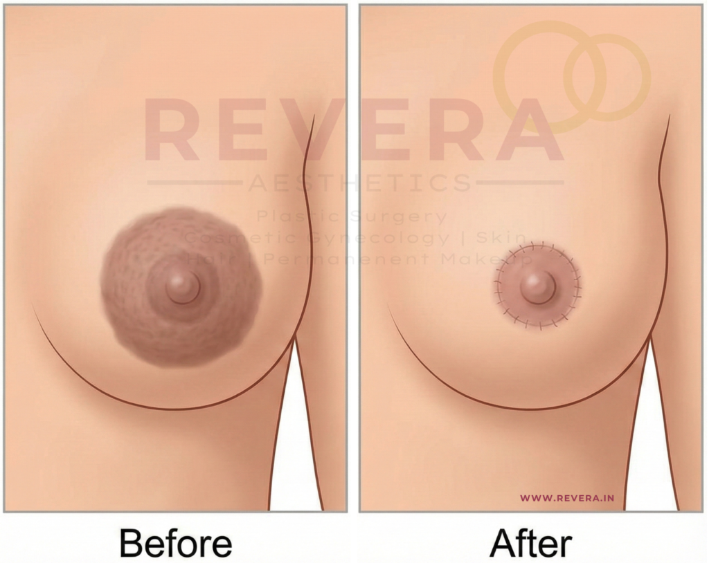
Breast reduction surgery is not just about making the breast smaller; it is about reshaping it to look proportional and centering the nipple on the new mound.

Most standard breast reduction techniques (like the popular Superomedial Pedicle) assume the nipple is starting from a standard sagging position. But what happens when a patient’s nipples are naturally positioned medially—meaning they sit closer to the cleavage or “point inward”?

In these cases, using standard techniques can be mechanically difficult. Trying to move an inward-facing nipple into a central position can restrict the blood supply or create tension, limiting how perfect the final result can be.

The Solution: The Superolateral Pedicle (SLP)

A new study published in Plastic and Reconstructive Surgery (August 2025) highlights a specific surgical approach designed exactly for this anatomy: the Superolateral Pedicle (SLP).

How It Works

In breast reduction, the nipple is kept alive on a “pedicle”—a bridge of tissue that preserves blood vessels and nerves.

  • Standard Way (Superomedial): The tissue bridge is usually kept on the inner/top side.
  • The SLP Way: For patients with medial nipples, the surgeon keeps the tissue bridge on the outer/top side (Superolateral).

By anchoring the nipple from the outside, the surgeon can more easily swing and rotate the nipple into the perfect central position without fighting the breast’s natural tissue resistance.

Is It Safe? The Research Say Yes

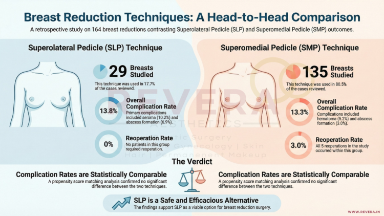
Researchers from MedStar Georgetown University Hospital in Washington, DC, reviewed 164 breast reductions to compare the safety of this specialized SLP technique against the standard method.

The Findings:

  • Comparable Safety: The complication rates were nearly identical between the two groups (13.8% for SLP vs. 13.3% for standard), proving that this technique is just as safe as the traditional method.
  • Effective Reduction: The technique worked well for significant reductions, with an average tissue removal of over 700g.
  • No Re-operations: In this specific study group, zero patients in the SLP group required a return to the operating room for complications, compared to 5 cases in the standard group.

Why This Matters For You

Anatomy is unique. If you have noticed that your nipples sit closer to your breastbone or point inward, standard techniques might not offer you the best aesthetic result. This research confirms that your surgeon has a validated, safe “tool in the toolkit” to correct medially positioned nipples and achieve a beautiful, centered look.


Frequently Asked Questions (FAQ)

Q: How do I know if I have “medially positioned” nipples?

A: If your nipples seem to sit closer to your cleavage rather than the center of your breast mound, or if they point inward towards each other, you likely have medial positioning. Your surgeon will assess this during your consultation.

Q: Does this technique leave different scars?

A: generally, no. The Superolateral Pedicle refers to the internal tissue handling. The external scars usually follow the standard “Wise Pattern” (Anchor) or Vertical (Lollipop) shape, just like a regular breast reduction.

Q: Is the recovery harder with this technique?

A: According to the study, complications such as wound healing issues (dehiscence) or fluid collection (seroma) were comparable to the standard technique, suggesting the recovery process is very similar.

Q: Can I still breastfeed with this technique?

A: Like the standard Superomedial technique, the SLP preserves a bridge of tissue carrying blood and nerve supply to the nipple. While breastfeeding can never be guaranteed after reduction surgery, techniques that preserve the pedicle generally offer a better chance than those that do not.


Reference

Lava, Christian X. MS; Li, Karen R. BBA; Episalla, Nicole C. MD; Snee, Isabel A. BS; Bell, Alice C. BA; Fan, Kenneth L. MD; Jabbour, Samer F. MD. “Superolateral Pedicle Breast Reduction for Patients with Medially Positioned Nipple-Areola Complexes.” Plastic and Reconstructive Surgery 156(2):p 174e-182e, August 2025.

Infographic on 2022 Evidence Based Safety Guidelines on Reduction Mammaplasty
Written by revera-admin

The New Gold Standard: What the Latest Guidelines (2022) Say About Breast Reduction

Setting the Standard for Safety and Results

Breast reduction surgery (reduction mammaplasty) is one of the most life-changing procedures in plastic surgery, performed on over 100,000 patients annually. To ensure patients receive the safest and most effective care, the American Society of Plastic Surgeons (ASPS) convened a multidisciplinary work group to update their clinical practice guidelines.

Published in 2022, these guidelines reviewed thousands of studies to determine what truly works. Here is what the new evidence means for you as a patient.

1. It Is About Your Symptoms, Not the Scale

For years, insurance companies often demanded a specific weight of tissue be removed (e.g., 500g or 1000g) to qualify for coverage. The new guidelines challenge this outdated metric.

The ASPS now strongly recommends that surgery be offered as first-line therapy based on symptoms, not resection weight.

  • The Evidence: Studies show that relief from back pain, neck pain, and bra strap grooving is not correlated with the amount of tissue removed.
  • The Takeaway: If you have multiple physical symptoms (pain, rashes, grooving) that aren’t fixed by non-surgical methods, you are a candidate for surgery, regardless of whether you need a “small” or “large” reduction6666.

2. Drains Are No Longer Routine

One of the most dreaded parts of recovery for many patients is the use of surgical drains (tubes sticking out of the incision to collect fluid).

The guidelines bring good news: Plastic surgeons should not routinely use intraoperative drains for breast reduction patients.

  • Why? High-quality evidence shows no significant difference in complication rates (like hematomas) between patients with drains and those without.
  • The Benefit: avoiding drains means less discomfort during removal, lower costs, and less scarring.

3. Pain Management Has Evolved (Less Narcotics)

The modern approach to breast reduction focuses on multimodal pain management to reduce the need for strong opioids (narcotics).

  • Local Anesthesia: The guidelines strongly recommend administering local anesthetic (numbing medication like lidocaine or bupivacaine) at the surgical site. This significantly improves pain scores immediately after surgery and reduces the time spent in the recovery room.
  • Non-Narcotic Strategies: Surgeons are encouraged to use non-opioid medications (such as Acetaminophen or NSAIDs) to manage pain safely.

4. Technique: The Pedicle Choice

The “pedicle” is the bridge of tissue that keeps your nipple alive and sensitive during the lift and reduction. The guidelines reviewed the two most common techniques:

  1. Inferior Pedicle: The most commonly used, reliable for preserving blood supply.
  2. Superomedial Pedicle: Preserves upper-pole fullness and avoids a long transverse scar.

The verdict? Both techniques are acceptable and effective. There is no significant difference in major complications between them, so your surgeon can choose the method best suited to your specific anatomy.

5. Important Risk Factors

To ensure safety, the guidelines identified specific factors that may increase the risk of complications. Patients should be counseled if they:

  • Are older than 50 years.
  • Have a Body Mass Index (BMI) greater than 35.
  • Use chronic corticosteroids.

Additionally, there is a strong recommendation regarding Nicotine: Patients identified as nicotine users should be referred to cessation programs and encouraged to stop smoking before surgery. Smoking significantly increases the risk of wound healing problems and infection.

6. Antibiotics and Pathology

  • Antibiotics: Extended courses of antibiotics after you go home are generally not recommended. A single dose given before surgery (within 1 hour of incision) is sufficient to prevent infection without causing antibiotic resistance.
  • Pathology: It is recommended that all breast tissue removed during the surgery be sent to the lab for evaluation to check for any hidden abnormal cells or high-risk lesions.

Frequently Asked Questions (FAQ)

Q: Do I have to try physical therapy before surgery?

A: The guidelines state that reduction mammaplasty should be offered as first-line therapy over non-operative treatments. There is no evidence that non-operative management (like special bras or therapy) provides effective long-term relief for breast hypertrophy.

Q: Will I have drains?

A: According to the 2022 guidelines, routine use of drains is not supported by evidence. However, exceptions may be made for specific high-risk cases or if liposuction is also performed.

Q: Does the “Pedicle” technique affect breastfeeding?

A: The guidelines noted that techniques preserving the subareolar parenchyma (the tissue under the nipple) increase the likelihood of breastfeeding success, but more research is needed to compare specific techniques directly.


Reference

Perdikis, Galen M.D.; Dillingham, Claire D.O.; et al. “Evidence-Based Clinical Practice Guideline: Revision: Reduction Mammaplasty.” Plastic and Reconstructive Surgery 149(3):p 392e-409e, March 2022.

Infographic showing Fully Awake Breast Surgery
Written by revera-admin

Awake Breast Reduction: Is General Anesthesia Necessary?

A Revolutionary Approach to Breast Surgery

For many women considering breast reduction surgery, the fear is not necessarily of the procedure itself, but of “going under.” General anesthesia, while generally safe, comes with side effects like nausea, grogginess, and a longer recovery time.

However, a study published in Plastic and Reconstructive Surgery presents a compelling alternative: Fully Awake Breast Reduction.

Authors Dr. Simon Filson, Dr. Danielle Yarhi, and Dr. Yitzhak Ramon from Haifa, Israel, successfully performed breast reductions on 25 patients who were awake, communicative, and able to move during the surgery—all without feeling pain.

How Is It Possible?

The secret lies in Thoracic Epidural Anesthesia.

Unlike general anesthesia, which puts the entire body to sleep and requires a breathing tube, a thoracic epidural numbs only the specific area of the body being operated on (the chest). The patient breathes on their own and remains conscious.

The “Awake” Cocktail: Anesthetics and Sedatives Used

To ensure the patient was comfortable, relaxed, and pain-free, the surgical team utilized a specific combination of oral medications and local anesthetics.

According to the study and the accompanying discussion by Dr. Donald Lalonde, the specific protocol included:

1. The “Sedative Cocktail”

Before the procedure began, patients were given these agents to induce relaxation and prevent pain or nausea:

  • Oxazepam : A benzodiazepine used to reduce anxiety.
  • Dipyrone : A strong analgesic (pain reliever).
  • Promethazine : An antihistamine that provides sedation and prevents nausea.

2. The Anesthetic Block (The Numbing Agents)

To perform the epidural block itself, the anesthesiologist used:

  • Lidocaine: Injected under the skin to numb the needle entry point.
  • Ropivacaine : Injected into the epidural space to provide the actual sensory block for the surgery.

Why Being Awake Changes Everything

The benefits of this technique went far beyond just avoiding general anesthesia. Because the patients were awake, they could actively cooperate during the surgery.

  • The “Sit-Up” Test: Patients could sit up on the operating table, allowing the surgeon to check the symmetry and shape of the breasts against gravity in real-time. The patient could even look and give their opinion before the surgery was finished!.
  • Faster Recovery: Patients were able to get off the operating table and walk to the recovery room immediately after surgery.
  • No “Hangover”: There were zero reports of nausea or vomiting, a common side effect of general anesthesia.

Expert Debate: “Sedated” vs. “Wide Awake”

This technique has sparked interesting conversations among experts. Dr. Donald Lalonde, a Canadian expert in “Wide-Awake Surgery,” applauded the move away from general anesthesia but noted that because sedatives (Oxazepam and Promethazine) were used, the patients were technically “sedated” rather than “wide awake”.

Dr. Lalonde suggested that in the future, surgeons might be able to skip the sedatives entirely and use Lidocaine with Epinephrine for the block, making the procedure truly “wide awake” and eliminating the risks associated with sedatives.

The Future of Surgery

Whether using mild sedation or pure local anesthesia, the trend is clear: surgery is becoming safer, lighter, and more patient-focused. If you are considering breast reduction but fear general anesthesia, this study proves that effective, comfortable alternatives exist.


Reference

Written by revera-admin

Finally, a Solution for Nipple Reshaping Without the Visible Scars

The Hidden Insecurity

While breast augmentation and lifts are commonly discussed, many patients struggle silently with insecurities regarding the nipple-areola complex. Whether due to genetics, hormonal changes, or the aftermath of breastfeeding, common complaints include:

  • Large Areolae: An areola that feels disproportionately large for the breast.
  • Undefined Contours: A lack of crisp definition between the nipple and the surrounding skin.
  • Herniated Tissue: Often called “puffy nipples,” where glandular tissue pushes out, creating a dome-like appearance.

For years, patients have hesitated to fix these issues for one main reason: The fear of scarring.

Usual Scars

The Old Trade-Off: Shape vs. Scars

Historically, correcting the shape of the nipple or reducing the size of the areola came with a compromise. Previous techniques, such as the “petal pattern method,” were effective at contouring but often limited in how much they could reduce the areola size.

More importantly, these older methods frequently resulted in visible scars from the front. For a patient seeking aesthetic improvement, trading a shape issue for a visible scar was often a difficult choice.

The Innovation: The “Crown-Shape” Method

A breakthrough study published in Plastic and Reconstructive Surgery (June 2025) introduces a refined solution: the Crown-Shape Debulking Method.

Developed by Dr. Ju Young Go and Dr. Won Lee from leading clinics in Seoul and Anyang, South Korea, this technique is a “single-stage” procedure designed to reshape the nipple without leaving obvious marks.

How It Works

The “Crown-Shape” technique is an advanced evolution of previous methods. It involves:

  1. Debulking: Removing the herniated or excess glandular tissue that causes puffiness.
  2. Structuring: Using a specific “crown” pattern to tighten the skin and define the nipple.
  3. Hiding the Evidence: The key innovation is that it allows for significant reduction and reshaping “without visible scars in the frontal areolar area”.

Why This Is a Game-Changer

This new Korean technique addresses the limitations of the past. It offers greater control during surgery to ensure the new shape is aesthetically pleasing while minimizing the risk of the problem recurring (recurrence).

Key Benefits:

  • Scar-Free Frontal View: No distracting scar lines on the face of the areola.
  • Effective Reduction: Can handle larger reductions than previous methods.
  • Better Definition: Creates a distinct, attractive nipple contour.

Is This Right for You?

If you have been bothered by “puffy” nipples or large areolas but have avoided surgery because you didn’t want scars, the Crown-Shape Debulking method might be the answer. This technique allows for a natural look that stands up to close inspection.


Frequently Asked Questions (FAQ)

Q: What specific problems does this fix?

A: This technique is designed for patients with large areolae, undefined nipple contours, or herniation of glandular tissue (bulging/puffy nipples) around the areola.

Q: How is this different from the “Petal Pattern” method?

A: The “Petal Pattern” was a previous technique that was good for contouring but had limitations in how much it could reduce the areola size. Furthermore, the Petal Pattern often left visible scars when viewed from the front. The Crown-Shape method improves upon this by allowing for better reduction without the visible frontal scars.

Q: Is there really no scar?

A: All surgery involves incisions, but this technique is described as “scar-free” in the frontal areolar area. This means the incisions are strategically placed and hidden so that when looking at the breast directly, there are no obvious surgical markings.


Reference

Go, Ju Young MD, PhD; Lee, Won MD, PhD. “Scar-Free Nipple and Areola Contouring: A Crown-shape Debulking Method for Enhanced Aesthetic Outcomes.” Plastic and Reconstructive Surgery. June 20, 2025. Seoul and Anyang, Korea.


Image depicts infographic of Nipple Sparing Inferior Flap Mammaplasty, the time taken, amount of tissue removed and Blood loss
Written by revera-admin

Relief for Massive Breasts: A Safer, Faster Technique for High-Risk Patients

When Breast Reduction is a Medical Necessity

For many women, breast reduction is about comfort and confidence. But for women with Gigantomastia (massive breasts), the condition is a severe medical burden.

Patients with massive breasts often suffer from debilitating back pain, skin infections, and significant mobility issues. Even more concerning, the sheer weight of the tissue can exacerbate pre-existing cardiac (heart) and respiratory (lung) problems.

For these patients, surgery is not a luxury—it is a necessity. However, because these patients often battle obesity or heart conditions, undergoing a long surgical procedure can be risky.

The Challenge: The 4-Hour Marathon

The most popular traditional methods for breast reduction (such as the McKissock or Robbins techniques) rely on complex internal sculpting. While effective, these surgeries typically require 3 to 4 hours of operative time.

For a patient with heart or lung issues, being under general anesthesia for four hours can be dangerous. Consequently, some women feel forced to consider a Total Mastectomy (complete removal of the breast) just to get relief, or they avoid surgery altogether.

The Solution: Nipple-Bearing Inferior Flap Mammaplasty

A technique published in Plastic and Reconstructive Surgery offers a powerful solution specifically designed for massive weight reduction with maximum safety.

Developed by a surgical team in Preston, England, this technique focuses on speed and blood supply safety.

How It Works

Unlike complex sculpting methods that require separating the breast tissue from the chest wall (“undermining”), this technique leaves the base of the breast undisturbed.

  1. Wide Base: The surgeon creates a wide, supportive base of tissue (the “inferior flap”) that keeps the nipple attached to its natural blood supply.
  2. No Grafting Needed: Unlike some rapid reduction techniques that cut the nipple off and sew it back on as a skin graft, this method keeps the nipple connected, preserving its vitality.
  3. Efficient Removal: A wedge of tissue is removed down to the pectoral fascia, and the remaining flaps are brought together securely.

The Results: Faster and Safer

The study followed patients with massive breasts and significant medical problems who underwent this specific procedure. The results were transformative:

  • Drastically Reduced Surgery Time: The average operation took only 88 minutes (compared to the standard 3–4 hours).
  • Massive Weight Loss: The average tissue removal was 2.76 kg (over 6 lbs) per patient.
  • Safety: There was minimal blood loss (less than 500 mL on average).
  • Nipple Survival: Because the nipple was carried on a wide, robust flap, it remained well-vascularized with no complications in the study group.

Is This Right For You?

This technique is ideal for women who:

  • Have extremely large breasts causing medical distress.
  • Have been told they are “high risk” for long surgeries due to obesity or heart/lung conditions.
  • Want significant size reduction without resorting to a total mastectomy.

We believe that health and mobility should never be out of reach. If you are suffering from the weight of massive breasts, contact us to discuss if this rapid-reduction technique is an option for you.


Frequently Asked Questions (FAQ)

Q: Will I lose my nipple sensation? A: This technique is a “Nipple-Bearing” procedure. Unlike a “Free Nipple Graft” (where the nipple is completely detached), this method maintains a wide bridge of tissue connecting the nipple to the body’s blood and nerve supply, which helps preserve the nipple-areola complex.

Q: How are the scars placed? A: The incision design usually results in an inverted-U or inverted-L shape on the lower breast. While scarring is inevitable in reduction surgery, the primary goal of this specific technique is massive volume reduction to improve heart and lung health.

Q: Why is a shorter surgery better? A: For patients with pre-existing medical conditions (like high blood pressure, asthma, or obesity), prolonged anesthesia increases the risk of complications such as clots (DVT) or respiratory distress. Reducing the time from 4 hours to roughly 90 minutes significantly lowers this risk.


Reference

Alvi, R. F.R.C.S.I.; Jaffe, W. F.R.C.S.; Laitung, J. K. G. Ch.M., F.R.C.S.Ed.. “Nipple-Bearing Inferior Flap Mammaplasty: A New Technique for Reducing Massive Breasts.” Plastic and Reconstructive Surgery 101(1):p 174-176, January 1998.

Written by revera-admin

More Than Just Shape: Restoring Feeling After Breast Reconstruction

The “Numb” Reality of Mastectomy

For millions of women undergoing mastectomy (breast removal) for cancer treatment or prevention, the primary focus is survival. The secondary focus is often reconstruction—restoring the physical appearance of the breast.

However, there is a “silent” side effect that is rarely discussed but deeply felt: Numbness.

During a mastectomy, the sensory nerves that provide feeling to the breast skin and nipple are often cut.

A Little Background on Anatomy

To understand why numbness occurs, we must look at how the breast receives feeling. The sensory innervation to the breast originates from the medial and lateral cutaneous branches of the third to fifth intercostal nerves.Image of thoracic dermatomes and intercostal nerve distribution

Shutterstock

  • The Path of the Nerves: The third, fourth, and fifth intercostal nerves give off lateral cutaneous branches that pierce the chest wall (near the mid-axillary line, or the side of your rib cage). These branches divide into anterior and posterior parts, supplying the skin of the side and front of the chest.
  • The Anterior Branches: The anterior cutaneous branches (the terminal parts of these nerves) supply the skin on the chest wall toward the center (sternum).
    • The 3rd nerve covers the upper-mid chest.
    • The 4th nerve covers the central chest and the medial (inner) breast area – The Nipple Line.
    • The 5th nerve covers the lower-mid chest and the inframammary fold (where the breast meets the ribs).

These nerves provide sensory innervation (known as dermatomes) to specific chest and abdominal areas and are crucial for sensation and pain management (such as nerve blocks).

For years, women have accepted that their reconstructed breasts, while looking beautiful, would permanently feel numb—like “wearing a bra made of your own skin.”

Restoring sensation (Resensation) is possible. Surgeons can perform a “nerve transfer,” connecting a nerve from your chest wall to the nerves in the reconstructed breast.

So, why isn’t this done all the time?

One major hurdle has been the difficulty of finding the right donor nerve. The human body is complex, and searching for a tiny nerve (often only 2 millimeters wide) during a long surgery can be like finding a needle in a haystack.

The Breakthrough: A Roadmap for Sensation

A pivotal study published in Plastic and Reconstructive Surgery has provided surgeons with a reliable “treasure map” to find these elusive nerves.

Research conducted by Dr. Rebecca Knackstedt and Dr. Risal Djohan (along with their team in Cleveland and Toledo, Ohio), utilized precise anatomical studies to identify the exact hiding place of the nerve responsible for breast sensation.

What They Found

The researchers discovered that the Lateral Intercostal Branch (the nerve key to breast feeling) is located in a highly predictable spot:

  • It almost always exits from under the 4th Rib.
  • It sits consistently near the edge of the Pectoralis Minor muscle.
  • It travels safely underneath the thoracodorsal vessels (major blood vessels in the armpit area).

Why This Matters for Your Surgery

This study transforms a “search mission” into a precise procedure. Because surgeons now know exactly where to look, we can locate the nerve with much greater accuracy. The study identified the nerve’s location as:

  • 10 to 15 cm from the sternum (breastbone).
  • 8 to 16 cm from the mid-clavicular line.
  • Near the lateral border of the armpit muscle (Pectoralis minor) or within 2 cm from it.

By using these precise coordinates, surgeons can:

  1. Locate the nerve quickly, reducing surgery time.
  2. Preserve the nerve more effectively.
  3. Perform Nerve Allografts: Connect this sensation-carrying nerve to your reconstructed breast tissue using a nerve graft.

Moving Beyond “Looking Normal”

We believe that feeling whole means more than just looking in the mirror; it’s about feeling a hug, noticing a change in temperature, and reclaiming your body’s sensation.

Thanks to anatomical breakthroughs like this study from Ohio, Breast Neurotization (nerve repair) is becoming a more standard and successful part of breast reconstruction.


Frequently Asked Questions (FAQ)

Q: If I have this procedure, will my sensation be 100% normal?

A: “Normal” is a strong word. Nerve regeneration is slow and complex. Most patients do not regain perfect, pre-surgery sensitivity. However, the goal is to transition from “numbness” to “protective sensation” (feeling touch and pressure) and, in many cases, erogenous sensation. It is a vast improvement over having no feeling at all.

Q: Does this add time to the surgery?

A: Yes, nerve repair does add some time to the reconstruction surgery. However, thanks to the “mapping” provided by this research, the time taken to find the nerve is significantly reduced, making the addition of nerve repair much more feasible.

Q: Can this be done if I had a mastectomy years ago?

A: Breast reinnervation is most successful when performed at the same time as the mastectomy (Immediate Reconstruction). Doing it years later is much more difficult because the nerve endings may have scar tissue or have become dormant. However, it is always worth discussing with your surgeon.

Q: Is this only for implant reconstruction or flap reconstruction?

A: Nerve grafts can be used in both. In DIEP Flap (using your own tissue), surgeons connect the chest nerve to the nerve in the tummy tissue. In Implant reconstruction, the nerve is connected to the remaining skin or nipple nerves.


Reference

Knackstedt, Rebecca M.D., Ph.D.; Gatherwright, James M.D.; Cakmakoglu, Cagri M.D.; Djohan, Michelle M.S.; Djohan, Risal M.D. “Predictable Location of Breast Sensory Nerves for Breast Reinnervation.” Plastic and Reconstructive Surgery. February 2019. Cleveland Clinic & University of Toledo, Ohio.

Written by revera-admin

Thinking About a Second Breast Reduction? New Research Makes “Revisions” Safer Than Ever

When One Surgery Isn’t Enough

Breast reduction surgery typically has one of the highest satisfaction rates in cosmetic medicine. However, bodies change. Due to weight fluctuations, hormonal shifts, or pregnancy, breast tissue can sometimes regrow, or gravity may cause sagging to return years after the initial procedure.

Many women find themselves wanting a Secondary Breast Reduction (a revision surgery) but hesitate due to safety concerns.

Historically, performing a reduction on a breast that has already been operated on was considered risky. The main fear? Compromising the blood supply to the nipple.

The “Unknown Pedicle” Problem

In a primary breast reduction, the surgeon creates a “pedicle”—a bridge of tissue that keeps the nipple and areola attached to their blood and nerve supply while the surrounding tissue is removed.

The challenge with revision surgery is that the new surgeon often doesn’t know which technique the previous surgeon used. If they cut into the old “lifeline” by mistake, it can lead to Nipple-Areola Complex (NAC) Necrosis (loss of the nipple tissue).

The Austrian Solution: A Triple-Safety Technique

A new prospective study published in April 2025 in the Plastic and Reconstructive Surgery journal offers a reassuring solution.

A team of researchers from Linz and Innsbruck, Austria, led by Dr. Sandra Feldler and Dr. Manfred Schmidt, has developed a “Modified McKissock Technique” specifically designed for these complex revision cases.

How It Works

The classic “McKissock” technique uses a vertical bipedicle (a bridge with a top and bottom attachment) to supply blood to the nipple.

The Austrian team modified this by adding a third component: a Central Pedicle.

  • Superior Pedicle (Top)
  • Inferior Pedicle (Bottom)
  • Central Pedicle (Middle)

Think of it as adding an extra emergency power line. Even if the surgeon doesn’t know exactly how the first surgery was performed, this “triple-threat” approach ensures the nipple retains a robust blood supply from multiple directions.

The Results: 100% Safety Record

The study followed 25 breast revisions using this new technique. The results were remarkably positive:

  • Zero Necrosis: There were no cases of nipple loss or tissue death.
  • Significant Reduction: The average patient had roughly 300g of tissue removed per breast.
  • High Satisfaction: 84.6% of patients rated their aesthetic appearance as “excellent” after the surgery.

Why This Matters For You

If you have been told that a second breast reduction is “too risky” or that you aren’t a candidate because your previous surgical records are lost, this research changes the conversation.

This modified technique allows surgeons to navigate the “unknowns” of your previous surgery with a safety net, ensuring you can achieve the smaller, lifted shape you desire without compromising your safety.


Frequently Asked Questions (FAQ)

Q: Why do breasts get big again after a reduction?

A: While the fat and glandular tissue removed during surgery is gone forever, the remaining cells can expand. Weight gain, pregnancy, menopause, and certain hormonal medications can stimulate the remaining breast tissue to grow.

Q: Is a revision recovery harder than the first time?

A: Surprisingly, many patients find the recovery similar or even slightly easier, as less tissue is usually removed compared to the first massive reduction. However, strict adherence to post-op care is vital to protect the blood supply.

Q: Does this technique leave more scars?

A: This technique generally utilizes the “inverted-T” or “anchor” scar pattern. Since most primary breast reductions also use this pattern, the surgeon simply goes through the old scar lines, meaning you likely won’t have new scars, just refreshed ones.

Q: Can I breastfeed after a secondary reduction?

A: Breastfeeding after a primary reduction is already difficult (about 50% success rate). A secondary reduction involves further manipulation of the milk ducts. While the nipple is kept alive and sensitive, the ability to breastfeed is unlikely after a second procedure.


Reference

Feldler, Sandra MD; Zaussinger, Maximilian MD; Ehebruster, Gudrun MD; Bachleitner, Kathrin MD; Steinkellner, Theresia MD; Schmidt, Manfred MD. “Modified McKissock Technique for Secondary Breast Reduction: A Prospective Study on Safety and Surgical and Aesthetic Outcomes.” Plastic and Reconstructive Surgery. April 2025. Linz and Innsbruck, Austria.


Written by revera-admin

Natural Volume Without Implants: A New Brazilian Technique for Restoring Breast Volume in Breast Lift & Breast Reduction

The Quest for the Perfect Profile

For many women considering a Mastopexy (Breast Lift) or Reduction Mammoplasty, the goal is simple: a lifted, youthful shape and a more manageable size.

However, there has always been a surgical dilemma. Traditional lifts are excellent at removing excess skin and reshaping the breast, but they often struggle to maintain “upper pole projection”—that desirable fullness at the top of the breast (the décolletage). Over time, gravity can cause the breast tissue to settle, sometimes leaving the upper breast looking flat.

Historically, the solution to this was using silicone implants to fill that upper volume. But what if you don’t want implants?

The “Implant-Free” Movement

While implants remain a popular choice, many of our patients are seeking natural alternatives. Some wish to avoid the maintenance of implants, while others are concerned about rare but documented risks associated with silicone, such as Breast Implant-Associated Anaplastic Large-Cell Lymphoma (BIA-ALCL) or autoimmune symptoms.

If you desire fullness and lift but want to stay 100% natural, a groundbreaking new technique known as Triple-Plane Autologous Fat Grafting might be the answer.

Innovative Research from Brazil

We pride ourselves on staying up-to-date with global advancements in aesthetic medicine. This specific technique comes from a cutting-edge study titled “Breast codes: triple plane autologous fat grafting,” published in August 2025 in the prestigious Plastic and Reconstructive Surgery journal.

The study was conducted by Dr. Ricardo T. Nóra, a plastic surgeon based in Sinop, Mato Grosso, Brazil, in collaboration with Dr. Lydia M. Ferreira from the Division of Plastic Surgery at the Federal University of São Paulo, Brazil.

Brazil has long been recognized as a world leader in cosmetic surgery innovation. In this study, Dr. Nóra and Dr. Ferreira standardized a method to enhance breast shape without foreign bodies, addressing the limitations of previous techniques.

What is the “Triple-Plane” Technique?

Autologous Fat Grafting involves taking fat from an area where you have a little extra (like the abdomen or thighs) and transferring it to the breasts.

The innovation introduced by Dr. Nóra and Dr. Ferreira is the “Triple-Plane” approach. Instead of randomly injecting fat, this technique systematically places fat in three specific layers (planes) to build a lasting structural foundation:

  1. Submuscular: Deep placement under the muscle for foundational volume.
  2. Intramuscular: Placement within the muscle for dynamic shaping.
  3. Subcutaneous: Placement just under the skin for smooth contouring and softness.

Why This Matters for You

This innovative approach addresses the “flat top” issue without requiring a foreign object in your body. By strategically layering the fat according to these new “Breast Codes,” surgeons can now create that coveted upper-breast fullness during a lift or reduction.

Key Benefits:

  • Natural Results: Uses your own tissue, so the look and feel are entirely you.
  • Safety: Eliminates the risks associated with long-term silicone implants.
  • Body Contouring: You get the added benefit of liposuction in the donor area.
  • Scientifically Backed: Based on rigorous surgical protocols developed by leading experts in Brazil.

Experience the Future of Breast Surgery

If you are looking for a natural, long-lasting enhancement, let’s discuss if this technique is right for your body goals. Contact our clinic today at +919866224871 to schedule a consultation. Chat with us on WhatsApp.

We are located in Hyderabad – Himayatnagar and Gachibowli – Kondapur.


Reference

Nóra, Ricardo T. M.D., MSc; Ferreira, Lydia M. M.D., PhD. “Breast codes: triple plane autologous fat grafting in mastopexies and reduction mammoplasties without implants.” Plastic and Reconstructive Surgery. August 13, 2025.

1 2 3 4